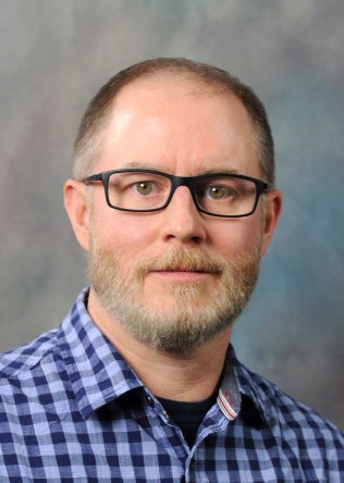
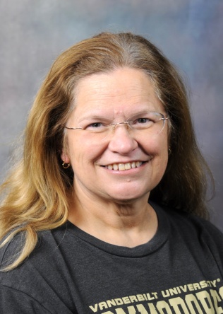
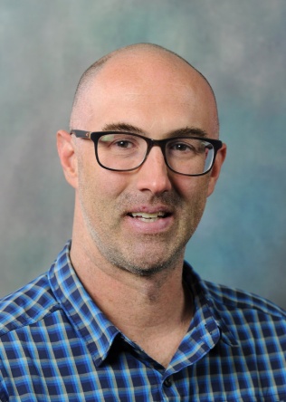
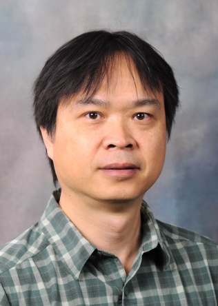
Noor Tantawy, Ph.D.

Mahammed
Noor
Tantawy
Ph.D
Research Associate Professor
Radiology & Radiological Sciences
Phone
(615)343-4795

Tantawy, M. N., T. E. Peterson, C. K. Jones, K. Johnson, J. M. Rook, P. J. Conn, R. M. Baldwin, M. S. Ansari, and R. M. Kessler, Impact of isoflurane anesthesia on D2 receptor occupancy by [18F]fallypride measured by microPET with a modified Logan plot: Synapse, [Epub ahead of print].

Tantawy, M. N., and T. E. Peterson, 2010 Apr, Simplified [18F]FDG image-derived input function using the left ventricle, liver, and one venous blood sample: Mol Imaging, v. 9, p. 76-86.

Tantawy, M. N., C. K. Jones, R. M. Baldwin, M. S. Ansari, P. J. Conn, R. M. Kessler, and T. E. Peterson, 2009, [18F]fallypride dopamine D2 receptor studies using delayed microPET scans and a modified Logan plot: Nucl Med Biol, v. 36, p. 931-40.

Manning, H. C., N. B. Merchant, A. C. Foutch, J. M. Virostko, S. K. Wyatt, C. Shah, E. T. McKinley, J. Xie, N. J. Mutic, M. K. Washington, B. LaFleur, M. N. Tantawy, T. E. Peterson, M. S. Ansari, R. M. Baldwin, M. L. Rothenberg, D. J. Bornhop, J. C. Gore, and R. J. Coffey, 2008, Molecular Imaging of Therapeutic Response to Epidermal Growth Factor Receptor Blockade in Colorectal Cancer: Clinical Cancer Research, v. 14, p. 7413-7422.

Hariri, G., Y. Zhang, A. Fu, Z. Han, M. Brechbiel, M. N. Tantawy, T. E. Peterson, R. Mernaugh, and D. Hallahan, 2008, Radiation-Guided P-Selectin Antibody Targeted to Lung Cancer: Annals of Biomedical Engineering, v. 36, p. 821-830.

n.tantawy@vumc.org

I do research using nuclear imaging modalities which include PET, SPECT, and CT. I collaborate with researchers from a wide range of diciplinaries.

Seth Smith, Ph.D.

Seth
Smith
Ph.D
Professor
Radiology & Radiological Sciences
Professor
Biomedical Engineering
Professor
Ophthalmology
Director of the Human Imaging Core
Phone
(615)421-8527

Reproducibility of tract-specific magnetization transfer and diffusion tensor imaging in the cervical spinal cord at 3 tesla. Smith SA, Jones CK, Gifford A, Belegu V, Chodkowski B, Farrell JA, Landman BA, Reich DS, Calabresi PA, McDonald JW, van Zijl PC.

Damage to the optic radiation in multiple sclerosis is associated with retinal injury and visual disability. Reich DS, Smith SA, Gordon-Lipkin EM, Ozturk A, Caffo BS, Balcer LJ, Calabresi PA. Arch Neurol. 2009 Aug;66(8):998-1006.

Direct saturation MRI: theory and application to imaging brain iron. Smith SA, Bulte JW, van Zijl PC. Magn Reson Med. 2009 Aug;62(2):384-93.

Sensorimotor dysfunction in multiple sclerosis and column-specific magnetization transfer-imaging abnormalities in the spinal cord. Zackowski KM, Smith SA, Reich DS, Gordon-Lipkin E, Chodkowski BA, Sambandan DR, Shteyman M, Bastian AJ, van Zijl PC, Calab

Quantitative magnetization transfer characteristics of the human cervical spinal cord in vivo: application to adrenomyeloneuropathy. Smith SA, Golay X, Fatemi A, Mahmood A, Raymond GV, Moser HW, van Zijl PC, Stanisz GJ. Magn Reson Med. 2009 Jan;61(1):22

seth.smith@vumc.org

My research is focused on translation of advanced, quantitative MRI methods to the human population at both high and low field strenghts.  Specifically, I am interested in developing advanced quantitative MRI methods to study under-represented structures of the human nervous system (optic nerve, spinal cord, and peripheral nerves) as they pertain to human disease.

We are currently involved in a number of research projects, all of which are targeted at understanding the pathophysiology of human disease through the development of advanced, quantitative MRI biomarkers for neurological diseases of the Brain, Spinal Cord, Optic Nerves, and Peripheral nerves.  Specifically, we are focused on ultra-high field (7T) CEST, Diffusion (conventional diffusion tensor and advanced diffusion weighted MRI), Magnetization Transfer, and quantiative T2.  

David Smith, Ph.D.

David
Smith
Ph.D
Assistant Professor
Radiology & Radiological Sciences, Physics & Astronomy, Electrical & Computer Engineering
Phone
(615)936-5166

Trajectory optimized NUFFT: Faster non?Cartesian MRI reconstruction through prior knowledge and parallel architectures. DS Smith, S Sengupta, SA Smith, EB Welch, 2019. Magnetic resonance in medicine 81 (3), 2064-2071. Trajectory optimized NUFFT: Faster non?Cartesian MRI reconstruction through prior knowledge and parallel architectures. DS Smith, S Sengupta, SA Smith, EB Welch, 2019. Magnetic resonance in medicine 81 (3), 2064-2071.

Dynamic Imaging of the Eye, Optic Nerve, and Extraocular Muscles With Golden Angle Radial MRI. S Sengupta, DS Smith, AK Smith, EB Welch, SA Smith, 2017. Investigative ophthalmology & visual science 58 (10), 4390-4398.

Continuously moving table MRI with golden angle radial sampling. S Sengupta, DS Smith, EB Welch, 2015. Magnetic resonance in medicine 74 (6), 1690-1697.

DCEMRI. jl: a fast, validated, open source toolkit for dynamic contrast enhanced MRI analysis. DS Smith, X Li, LR Arlinghaus, TE Yankeelov, EB Welch, 2015. PeerJ 3, e909.

Gamma-ray Exposure Rate Constants and Lead Shielding Data for over 1200 Radionuclides. DS Smith and M Stabin, 2010. Physics in Medicine and Biology, 102 (3), 271-291.

david.smith@vumc.org

Some of my current work includes cancer imaging, radiomics, data science, deep learning, parallel computing, optimization, applied math, physics, astronomy, and astrobiology.

Saikat Sengupta, Ph.D.

Saikat
Sengupta
Ph.D
Research Associate Professor
Radiology & Radiological Sciences

Sengupta S, Smith DS, Gifford A, Welch EB Whole-body Continuously Moving Table Fat-Water MRI with Dynamic B0 Shimming at 3 Tesla. Magn Reson Med. (In Press)

Sengupta S, Smith DS, Welch EB Continuously Moving Table MRI with Golden Angle Radial Sampling. Magn Reson Med. Oct 2014.

Sengupta S, Tadanki S, Gore JC, Welch EB Prospective real-time head motion correction using inductively coupled wireless NMR probes. Magn Reson Med. 2013 Nov 18.

Sengupta S, Welch EB, Zhao Y, Foxall D, Starewicz P, Anderson AW, Gore JC, Avison MJ. Dynamic B0 shimming at 7 Tesla. Magn Reson Imaging. 2011 May; 29(4): 483-96.

Sengupta S, Avison MJ, Gore JC, Welch EB. Software compensation of eddy current fields in multislice high order dynamic shimming. J Magn Reson. 2011 Jun; 210(2): 218-27.

saikat.sengupta@vumc.org

My interests lie in improving Magnetic Resonance Imaging performance by developing methods to minimize image artifacts and improve scanning efficiency on high field imaging systems. My research areas include real time shimming, continuously moving table imaging, motion correction and field monitoring.

My current work involves developing methods for rapid whole-body imaging using continuously moving table MRI methods. My past work has included realtime head motion correction using NMR probes for high resolution neuroimaging, dynamic B0 shimming at high field and developing models for prospective shim eddy current compensation.

Kurt Schilling, Ph.D.

Kurt
Schilling
Ph.D
Research Assistant Professor
Radiology & Radiological Sciences
kurt.g.schilling.1@vumc.org

My research is focused on image processing, biomedical modeling of magnetic resonance imaging (MRI) data, and mapping the human brain, with a focus on diffusion MRI data.

Dr. Schilling's research is focused on image processing, biomedical modeling of magnetic resonance imaging (MRI) data, and mapping the human brain, with a focus on diffusion MRI data

Baxter P. Rogers, Ph.D.

Baxter
P.
Rogers
Ph.D
Research Professor
Radiology & Radiological Sciences, Biomedical Engineering, Psychiatry
Phone
(615)343-4636

SB Katwal, JC Gore, R Marois, BP Rogers. Unsupervised spatiotemporal analysis of fMRI data using graph-based visualizations of self-organizing maps. IEEE Trans Biomed Eng 60(9):2472-83 (2013).  

BP Rogers, JM Sheffield, AS Luksik, S Heckers. Systematic error in hippocampal volume asymmetry measurement is minimal with a manual segmentation protocol. Frontiers in Neuroscience 6:179 (2012).

SB Katwal, JC Gore, JC Gatenby, BP Rogers. Measuring relative timings of brain activities using fMRI. Neuroimage 66:436-448 (2012).

ND Woodward, B Rogers, S Heckers. Functional resting-state networks are differentially affected in schizophrenia. Schizophr Res 130(1-3):86-93 (2011).

BP Rogers, VL Morgan, AT Newton, JC Gore. Assessing functional connectivity in the human brain by fMRI. Review. Magn Reson Imaging 25(10):1347-57 (2007).

baxter.rogers@vumc.org

I study the structure and function of the brain. I am particularly interested in methods of measuring the timing of functional MRI signals and the correlations between them, and in applications of these methods to the study of neurological and mental disorders.

I am currently involved in a series of studies of hippocampal structure and function in epilepsy and psychosis, using ultra high field MRI with new methods of measuring functional connectivity and statistical approaches appropriate for the large data sets involved. Recent methodological work is focused on understanding dynamic variation in functional connectivity over short time scales (minute to minute).

Wellington Pham, Ph.D.

Wellington
Pham
Ph.D
Professor
Radiology & Radiological Sciences
Professor
Biomedical Engineering
Phone
(615)936-7621
wellington.pham@vumc.org

Our group's research interests focus on the use of synthetic and colloidal chemistry in the development of novel molecular probes. Through our use of emerging imaging technologies, we foresee such probes being employed to investigate and define the mechanisms that lead to pathological diseases.

Our ongoing investigation emphasizes on cell therapy using a transgenic and preclinical mouse model. In addition, we have employed integrated nanotechnology-based platforms to achieve a multifunctional and multiplexed vaccine delivery system for cancer therapy. Aside from highlighting the contrast properties and carrier features available in nanotechnology, the overall goals of our studies are to (i) provide microanatomical and functional imaging feedback of the therapeutic process and (ii) realize an approach for longitudinal treatment and monitoring.     Our second project focuses on the development of a novel synthetic chemistry approach to the generation of probes intended specifically for imaging Alzheimer's disease (AD). The significance of this work lies in the development of a versatile vehicle which, after being loaded with imaging cargo, can be delivered to the brain. Furthermore, our laboratory currently maintains a clone of a double transgenic mouse model of AD to enable testing of these probes. Considering the tremendous contribution made by imaging in understanding the pathogenesis of AD, the results obtained through this research will have a strong influence on current efforts to find reliable biomarkers of this disease.

Todd Peterson, Ph.D.

Todd
Peterson
Ph.D
Professor
Radiology & Radiological Sciences
Director of Nuclear Imaging
Director of Radiochemistry
Master of Imaging Science Program Director
Phone
(615)322-2648

Lin A, Kupinski MA, Peterson TE, Shokouhi S, Johnson LC. Task-based Design of a Synthetic-collimator SPECT System Used for Small-animal Imaging. Medical Physics 2018;45(7):295202963 DOI: 10.1002/mp.12952

Johnson LC, Ovchinnikov O, Shokouhi S, Peterson TE. Development of a Germanium Small-Animal SPECT System. IEEE Transactions on Nuclear Science 2015;62(5):2036-2042 DOI: 10.1109/TNS.2015.2448673

Johnson LC, Shokouhi S, Peterson TE. Reducing multiplexing artifacts in multi-pinhole SPECT with a stacked silicon-germanium system: a simulation study. IEEE Transactions on Medical Imaging 2014;33(12):2342-2351 DOI: 10.1109/TMI.2014.2340251

Peterson TE and Furenlid LR. SPECT Detectors: the Anger Camera and beyond, Physics in Medicine and Biology 2011; 56:R145-R182

Tantawy MN, Peterson TE. Simplified [18F]FDG Image-Derived Input Function using the Left Ventricle, Liver, and One Venous Blood Sample. Molecular Imaging 2010;9(2):76-86

todd.e.peterson@vumc.org

My research has two major thrusts: 1.) the development of high-resolution, multi-pinhole SPECT capabilities, and 2.) the use of CT, SPECT, and PET in preclinical research studies across a wide range of applications.

We are developing novel semiconductor radiation detector technology for use in innovative multi-pinhole SPECT systems. 2.) We are involved in a number of collaborative projects in which we our role is to refine and validate image acquisition and analysis techniques to improve quantitative PET and SPECT capabilities.

Kristin O'Grady, Ph.D.

Kristin
O'Grady
Ph.D
Assistant Professor
Radiology & Radiological Sciences, Biomedical Engineering
Phone
(615)322-7209

K.G. Schilling, S. Fadnavis, J. Batson, M. Visagie, A.J.E. Combes, S. By, C.D. McKnight, F. Bagnato, E. Garyfallidis, B.A. Landman, S.A. Smith, & K.P. O'Grady. "Denoising of diffusion MRI in the cervical spinal cord - effects of denoising strategy and acquisition on intra-cord contrast, signal modeling, and feature conspicuity." NeuroImage: 266: 119826 (2023). Epub: December 18, 2022. PMID: 36543265. PMCID: PMC9843739.

A.J.E. Combes, M.A. Clarke, K.P. O'Grady, K.G. Schilling, & S.A. Smith. "Advanced spinal cord MRI in multiple sclerosis: Current techniques and future directions" NeuroImage: Clinical 36: 103244 (2022). PMID: 36306717. PMCID: PMC9668663.

A.J.E. Combes, K.P. O'Grady, B.P. Rogers, K.G. Schilling, R.D. Lawless, M. Visagie, D.C. Houston, L.E. Prock, S.D. Malone, S. Satish, A.A. Witt, C.D. McKnight, F. Bagnato, J.C. Gore, & S.A. Smith. "Functional connectivity in the dorsal network of the cervical spinal cord is correlated with diffusion tensor imaging indices in relapsing-remitting multiple sclerosis." NeuroImage: Clinical 35: 103127 (2022). PMID: 35917721. PMCID: PMC9421501.

K.P. O'Grady, S. Satish, Q.R. Owen, B.A. Box, F. Bagnato, A.J.E. Combes, S.R. Cook, H. Westervelt, H.R. Feiler, R.D. Lawless, A. Sarma, S.D. Malone, J.M. Ndolo, K. Yoon, R.D. Dortch, B.P. Rogers, & S.A. Smith. "Relaxation-compensated chemical exchange saturation transfer MRI in the brain at 7T: application in relapsing-remitting multiple sclerosis." Frontiers in Neurology - Applied Neuroimaging 13: 764690 (2022). Epub: February 25, 2022. PMID: 35299614. PMCID: PMC8923037.

K.P. O'Grady, A.N. Dula, B.D. Lyttle, L.M. Thompson, B.N. Conrad, B.A. Box, L.J. McKeithan, S. Pawate, F. Bagnato, B.A. Landman, P. Newhouse, & S.A. Smith. "Glutamate-sensitive imaging and evaluation of cognitive impairment in multiple sclerosis." Multiple Sclerosis Journal 25(12): 1580-1592 (2019). Epub: September 19, 2018. PMID: 30230400. PMCID: PMC6424643.

kristin.p.ogrady@vumc.org

I am interested in translating quantitative MR techniques to the clinic to better understand the pathological changes in the brain and spinal cord associated with neurological diseases such as multiple sclerosis.

My research is focused on developing quantitative MR methodologies for the brain and spinal cord with a current emphasis on improving thoracic and lumbar spinal cord MRI. This work is also part of an effort to improve our understanding of the effects of biological sex on pathological changes associated with neurological diseases such as multiple sclerosis.

Allen Newton, Ph.D.

Allen
Newton
Ph.D
Associate Professor
Radiology & Radiological Sciences, Pediatrics
allen.t.newton@vumc.org

I am interested in the development of new fMRI methods including advances in both image acquisition and image analysis. I am particularly interested in methods involving imaging at ultra high field (7T).

Currently, I have several parallel research paths I am pursuing. First, I am developing methods for pushing the spatial resolution of whole brain fMRI data. The goal of this project is to obtain images with the highest possible isotropic spatial resolution while minimizing geometric distortions of the images and maintaining reasonable temporal resolution. Second, I am developing methods for similarly pushing the temporal resolution of fMRI data while maintaining spatial resolutions similar to those typically used at lower field strengths. These acquisitions will be used for measuring cognitive latencies and validating methods for removing physiological noise.