What we can do for you
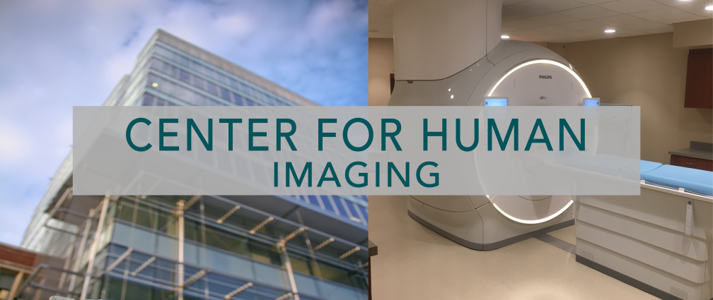
The Human Imaging Core (HIC) provides resources for structural and functional imaging and spectroscopy. Magnetic resonance imaging (MRI) and spectroscopy (MRS) are available on our two 3 Tesla full body scanners and a 7 Tesla full body scanner. In addition to data acquisition, we support MRI and MRS protocol development, functional MRI (fMRI) experimental design, subject preparation, structural and functional image analysis, and training on image analysis techniques and tools. Additional resources available include optical coherence tomography (OCT), Dual Energy X-ray Absorptiometry (DXA), PET/CT, and ultrasound.
Description
The Human Imaging Core is dedicated to the application of advanced and prototypical MRI methods to study specific organ systems of the human body such as: nervous (central and peripheral), muscular-skeletal, cardio-pulmonary, and integumentary systems. Additionally, many ongoing projects focus on development and validation of novel MRI biomarkers for the assessment of numerous diseases (e.g. cancer, multiple sclerosis, amyotrophic lateral sclerosis, epilepsy, concussion), conditions (cognitive impairment, learning disability), and normal function (activation of the human brain, sensitivity to pain). Specifically, our researchers are working on MRI methods sensitive to 1) tissue anisotropy, 2) tissue composition, 3) tissue metabolism, and 4) tissue function.
Information
Training
- User Guide
- VUIIS HIC Quick FAQ
- Resource calendars: Book It Lab
- Resource billing: iLab
- Data retrieval(VPN connection needed): gStudy
- Book It Lab User Training
General Resources
MRI Training
- MR User Training Session Registration
- MRI Zoom Recorded New User Orientation
- MRI Safety Training, Fall 2021
- User MRI Safety Screening
MRI Resources
- MRI worksheet
- MRI Incidental Finding Form
- MRI Safety Screening Form for Participants
- NHP MRI Form
- MRI Matching/Development Reservation Request
PET/CT
DXA
X-Ray and Ultrasound