Equipment | Contact | Description |
| 4.7T Small Animal MRI/MRS | Mark D. Does Daniel Colvin |
The 4.7T 31-cm bore Varian DirectDrive scanner is a fully broad-banded imaging/spectroscopy system equipped with actively shielded gradients (40 G/cm, rise times full amplitude of 130 μs), two independent transmit and one independent receive channels. Three transmit/receive quadrature imaging volume coils are available: 38 mm Litz coil (mouse), 63 mm birdcage coil(rat), and 160 mm birdcage coil (rabbit). System operation and animal prep support can be provided based on availability and need. The VUIIS provides computer access for data processing.
|
| 7.0T Small Animal MRI/MRS | Mark D. Does Daniel C. Colvin |
Three transmit/receive quadrature volume coils are available: 25 mm (mouse head), 38 mm (mouse body), and 72 mm (rat body), and the 72 mm coil can also be used with a 4-channel receive array suitable for rat head or mouse body. System operation and animal prep support can be provided based on availability and need.
|
| 9.4T Small Animal MRI/MRS | Mark D. Does Daniel C. Colvin |
The 9.4T 21-cm bore Bruker Avance NEO scanner is a fully broad-banded imaging/spectroscopy system equipped with actively shielded gradients (40 G/cm, rise times full amplitude of 135 μs), two independent 1H transmit/receive channels, a broadband transmit/receive channel, and four independent receive-only channels. Three transmit/receive quadrature imaging volume coils are available: 38mm Litz coil (mouse), 63mm birdcage coil (rat), and 85mm birdcage coil. A variety of X-nuclei surface coils are also available. System operation and animal prep support can be provided based on availability and need. The VUIIS provides computer access for data processing.
|
| 15.2T Small Animal MRI/MRS | Mark D. Does Daniel C. Colvin |
The 15.2T Bruker Biospec Avance III, 11cm horizontal bore scanner is a fully broadband imaging system equipped with 6 cm inner diameter actively shielded gradients (up to 100 G/cm, 107 μs rise time to max amplitude), and independent 1H and X nuclei transmit and receive channels. Three quadrature imaging coils are currently available: 35 mm volume birdcage coil (mouse), 35 mm volume birdcage coil for mouse and rat brain from Doty Scientific, Inc (Columbia, SC), and a closed cycle helium cryoprobe (quadrature surface coil) for high-resolution in vivo mouse imaging.
|
| Siemens Inveon PET/CT | Todd Peterson Noor Tantawy |
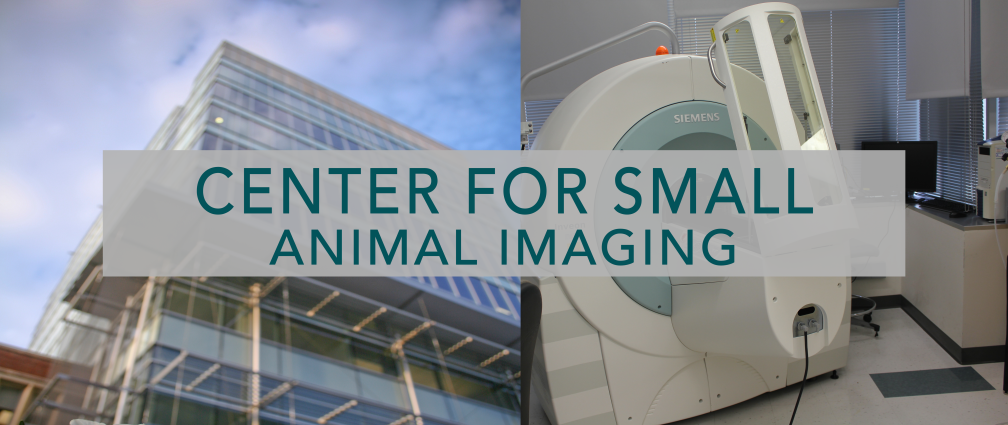
The Siemens Inveon multimodality PET/CT imaging scanner is capable of providing three dimensional CT and PET images of live mouse and rat as well as fixed samples. The system can deliver high resolution CT images (<10 µM for a field-of-view <20mm) with a maximum field-of-view of 80 mm X 50 mm (with a resolution of approximately 50 µM). The minimum resolution achievable with the PET scanner is approximately 1 mm (maximum field-of-view of 120 mm) with a high sensitivity in pico-Molar range.
|
| Bioscan NanoSPECT/CT | Todd Peterson Noor Tantawy |
The Bioscan NanoSPECT/CT system combines a single-photon emission computed tomography (SPECT) system with an x-ray CT. The SPECT system consists of four gamma cameras, each of which is outfitted with a nine-pinhole imaging aperture. Three different sets of pinhole apertures are available: one set for rat imaging (2.5 mm diameter pinholes), one set for high-sensitivity mouse imaging (1.4 mm diameter pinholes), and one set for high-resolution mouse imaging (1.0 mm diameter pinholes). The system is capable of acquiring dynamic images as well as dual-isotope imaging. All standard SPECT radionuclides can be accommodated (99mTc, 125I, 123I, 111In). The x-ray CT scanner and SPECT system are fully integrated and aligned such that co-registered CT images provide anatomical information to assist in interpretation and analysis of the SPECT images. Several radiotracers are routinely available from the VUMC radiopharmacy, including 99mTc-sestamibi, 99mTc-pertechnetate, 99mTc-MDP, and 99mTc-MAG3.
|
| Autoradiography | Noor Tantawy | Biospace Lab Micro Imager |
| Scanco vivaCT80 | Jeff Nyman |
The vivaCT80 is an X-ray CT scanner for in vivo imaging of small animals with a maximum diameter of 90 mm and isotropic image voxels as small as 5 um with a radiation dose low enough for repeated in vivo scans. This system is supported by the same analytical and visualization software as the Scanco specimen scanners.
|
| Scanco uCT40 and Scanco uCT50 | Jeff Nyman |
The Scanco uCT40 and uCT50 are x-ray CT scanners for obtaining and analyzing three-dimensional images of ex vivo specimens soft and hard tissues and materials. These state-of-the-art instruments are capable of producing images with voxel sizes as small as 6 um and 0.5 um, depending on specimen size, and can accommodate specimens as large as 36 mm or 48 mm in diameter, respectively. Both instruments are supported by cluster of seven HP Integrity servers for parallel processing on 42 CPU cores using a powerful suite of software designed by the manufacturer. The software suite contains several standardized and accepted methods for image reconstruction, VOI selection, segmentation, and analysis of structural, volumetric, and densitometric parameters. Additionally, we have extensive experience creating custom analytical algorithms within the Image Processing Language framework for a wide array of applications. Once created by our support team, these custom analyses can easily be executed by novice users through the Scanco Evaluation software. Visualization of three-dimensional renderings can be accomplished in the manufacturer’s software or images can be exported to a number of standard file formats for viewing in other commercially available software and/or coregistration with other imaging modalities. Users are trained VUIIS personnel.
|
| Faxitron XRAY | Jeff Nyman |
The Faxitron LX-60 X-ray system is utilized to produce highly detailed radiographs of small animals. The system’s large 12 cm x 12 cm camera offers exceptional 10 lp/mm spatial resolution in the contact mode. Magnification allows for spatial resolution of up to 60 lp/mm. The 20 μm nominal focal spot and additional geometric magnification provide ultra high-contrast and high spatial resolution images.
|
| CRI Maestro Optical | Jarrod True |
The CRI Maestro optical imaging system is designed primarily for in vivo fluorescence imaging. The Maestro is capable of background discrimination (autofluorescence) using spectral un-mixing techniques.
|
| IVIS Spectrum | Wellington Pham Jarrod True |
PerkinElmer IVIS Spectrum. The IVIS Spectrum offers an optimized set of 28 high efficiency filters spanning 430-850 nm and spectral un-mixing algorithms that let you take full advantage of bioluminescent and fluorescent reporters across the blue to near infrared wavelength region. It also offers single-view 3D tomography for both fluorescent and bioluminescent reporters that can be analyzed in an anatomical context using PerkinElmer's Digital Mouse Atlas or registered with their multimodality module to other tomographic technologies such as MR, CT or PET. For advanced fluorescence pre-clinical imaging, the IVIS Spectrum has the capability to use either trans-illumination (from the bottom) or epi-illumination (from the top) to illuminate in vivo fluorescent sources. 3D diffuse fluorescence tomography can be performed to determine source localization and concentration using the combination of structured light and trans illumination fluorescent images. The instrument is equipped with 10 narrow band excitation filters (30nm bandwidth) and 18 narrow band emission filters (20nm bandwidth) that assist in significantly reducing autofluorescence by the spectral scanning of filters and the use of spectral unmixing algorithms. In addition, the spectral unmixing tools allow the researcher to separate signals from multiple fluorescent reporters within the same animal.
|
| Visen FMT2500 | Jarrod True |
The FMT 2500 LX is a 3D fluorescence tomographic system with the ability to quantitate up to four fluorophores emitting 660, 700, 775 and 805nm wavelengths (excitation lights at 635, 670, 746 and 790nm, respectively). The emission signals are captured by a cooled-CCD and the data is processed using TrueQuantTM software. The algorithm takes into account tissue heterogeneity in reconstruction of the 3D image. FMT data can be co-registered with other imaging modalities based on the fiduciary marks on the imaging cassette that holds the subject. This is useful for many applications including oncology, inflammation and cardiovascular disease.
|
| Prep/Monitoring | Jarrod True |
All animal studies must have prior IACUC approval. Animal ventilators, anesthesia machines, and physiological monitoring systems are available for all imaging systems in the CSAI. Users may be billed for additional anesthetics, gases and animal prep supplies.
|

