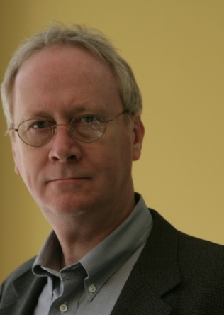
John Gore, Ph.D.

John
Gore
Ph.D
Distinguished University Professor
Radiology & Radiological Sciences, Biomedical Engineering, Molecular Physiology & Biophysics, Physics & Astronomy
Hertha Ramsey Cress Chair in Medicine
Director
Vanderbilt Institute of Imaging Science
(615)322-8357

Jeong, Ha-Kyu, John C Gore, and Adam W Anderson. "High-resolution Human Diffusion Tensor Imaging Using 2-D Navigated Multishot SENSE EPI at 7 T." Magnetic Resonance in Medicine, 69, no. 3 (March 1, 2013): 793"802.

Cobb, Jared Guthrie, Jingping Xie, and John C Gore. "Contributions of Chemical and Diffusive Exchange to T(1ρ) Dispersion." Magnetic Resonance in Medicine, (July 12, 2012).

Katwal, Santosh B, John C Gore, J Christopher Gatenby, and Baxter P Rogers. "Measuring Relative Timings of Brain Activities Using fMRI." NeuroImage 66C (October 27, 2012): 436"448.

Xu, Junzhong, Ke Li, R Adam Smith, John C Waterton, Ping Zhao, Heidi Chen, Mark D Does, H Charles Manning, and John C Gore. "Characterizing Tumor Response to Chemotherapy at Various Length Scales Using Temporal Diffusion Spectroscopy." PloS One 7, no. 7 (2012): e41714.

Chen, Limin, Arabinda Mishra, Allen T Newton, Victoria L Morgan, Elizabeth A Stringer, Baxter P Rogers, and John C Gore. "Fine-scale Functional Connectivity in Somatosensory Cortex Revealed by High-resolution fMRI." Magnetic Resonance Imaging 29, no. 10 (December 2011): 1330"1337

john.gore@vumc.org

I'm interested in the development and application of imaging science in medicine and biology, and the development of imaging science as a coherent discipline for trainees.

For almost 40 years my research has spanned different areas of imaging science - from basic physics of image formation, to understanding the factors that affect the signals used to make images, to applications that derive new types of information about biological systems, to image analysis and perception. Imaging of human subjects, non-human primates and small animals provides unique information on tissue structure and function, and is being applied in a variety of different applications in neuroscience, cancer research and studies of metabolism. A general theme of interest is to understand the physical and physiological factors that affect MRI signals and to use this knowledge to devise non-invasive imaging methods that provide new types of information, as well as for developing new applications of imaging. Fundamental studies of the basis of MRI contrast mechanisms such as proton relaxation, diffusion, and the BOLD effect in fMRI help interpret images used in numerous applications. A second major theme is the development of methods for studying human brain structure and function using MRI, especially at very high fields, and for integrating fMRI data with other imaging methods such as NIR and EEG. Several novel engineering solutions are being developed to address the technical challenges of human imaging at 7 Tesla, while other research to develop robust measurements of neurotransmitters and metabolism by multinuclear MRS is also being pursued. Applications of structural and functional MRI to the brain are performed in collaboration with investigators from psychology, psychiatry and other departments. A third major theme is the use of multi-modality imaging (MRI, PET, CT, optical and ultrasound) to study small animals, especially mouse models of human cancer and other genetically modified mice. New technologies being developed include acousto-optical imaging of fluorophores. Many projects also involve the development and application of advanced image analysis methods and computer algorithms.

Daniel Gochberg, Ph.D.

Daniel
Gochberg
Ph.D
Professor
Radiology & Radiological Sciences
daniel.gochberg@vumc.org

Our research focuses on the development, characterization, and application of MRI methods for quantifying tissue parameters, most importantly exchange between tissue water and macromolecules and metabolites. Such methods have their greatest application in imaging multiple sclerosis (MS).

We are currently working on developing and applying methods for measuring spin exchange, including developing new methods to detect tissue exchange sites (such as amides) using chemical exchange saturation transfer (CEST) and T1p. Also, we are applying quantitative magnetization transfer (qMT) methods to a rat model of multiple sclerosis in order to determine the method's sensitivity to myelin. This work may result in improved methods for research and clinical imaging.


Yurui Gao, Ph.D.

Yurui
Gao
Ph.D
Research Associate Professor
Biomedical Engineering
Phone
(615)343-8392

Gao Y, Choe AS, Li X, Stepniewska I, Anderson AW. Validation of DTI measures of primary motor area cortical connectivity. In Proceedings of the Joint Annual Meeting ISMRM-ESMRMB 2010, Stockholm, Sweden, 2010. (Abstract 117)

Choe AS, Gao Y, Stepniewska I, Li X, Ding Z, Anderson AW. Challenges of cortical connectivity measurements using MR tractography. In Proceedings of the Joint Annual Meeting ISMRM-ESMRMB 2010, Stockholm, Sweden, 2010. (Abstract 1665)

yurui.gao@vanderbilt.edu

I am interested in the relationship between diffusion magnetic resonance imaging and the distribution of axon fibers in brain white matter measured by light microscopy.

I am currently involved in the project focused on testing the ability of DTI tractograpy to investigate the cortical-cortical connectivity of the monkey brain. I will compare fiber distribution pattern calculated  by deterministic  algorithm to the histological fiber distribution determined by the stain of injected, intra-axonal tracer. 

Mark Does, Ph.D.

Mark
Does
Ph.D
Professor
Biomedical Engineering, Radiology & Radiological Sciences, Electrical Engineering
Director
Center for Small Animal Imaging
Phone
(615)322-8352

K. L. West, N. D. Kelm, R. P. Carson, M. D. Does, A Revised Model for Estimating g-ratio from MRI, Neuroimage, Vol 125:1155-1158, 2016.

N. D. Kelm, K. L. West, R. P. Carson, D. F. Gochberg, K C. Ess, M D. Does, Evaluation of Diffusion Kurtosis Imaging in Hypomyelinated Mouse Models, Neuroimage, 124(Pt A):612-26., 2016.

M. K. Manhard, R. A. Horch, D. F. Gochberg J. S. Nyman, M. D. Does, In Vivo Magnetic Resonance Imaging of Bound and Pore Water in Cortical Bone, Radiology, Oct;277(1):221-9, 2015.

C. L. Lankford, R. D. Dortch, M. D. Does, Fast T2 Mapping with Multiple Echo, Caesar cipher Acquisition and Model-Based Reconstruction, Magnetic Resonance in Medicine, 73(3):1065-74, 2015.

K. D. Harkins, M. D. Does, W. A. Grissom, An iterative method for predistortion of gradient waveforms, IEEE Transactions on Medical Imaging, Vol 33(8):1641 - 1647, 2014.

mark.does@vanderbilt.edu

We are interested in developing and applying MRI methods to quantitatively characterize various properties and/or compositions of tissue. To this end we develop models of NMR relaxation and water diffusion in tissue, develop and optimize MRI pulse sequences and associated technology, and experimentally investigate in vivo and ex vivo tissue models.

Current projects include: i) characterizing myelin content and thickness in normal and injured neural tissues using multi-exponential transverse relaxation, magnetization transfer, water diffusion, and ultra-short T2 methods; ii) characterizing fracture properties of cortical bone through T2-discriminated measures of bone collagen, collagen-bound water, and porosity; iii) characterizing edema and muscle fiber condition in models of skeletal muscle injury and disease using T2 and water diffusion; iv) investigating the effects of inter-compartmental water exchange on MRI measures of neural tissue, muscle, and tumors using relaxation-based exchange spectroscopy and SPECT imaging.

Zhaohua Ding, Ph.D.

Zhaouha
Ding
Ph.D
Research Professor
Electrical and Computer Engineering, Computer Science, Biomedical Engineering

My current projects include understanding fundamental structure-function relations in brain neural circuits, establishing mathematical models that characterize the dynamics of complex neural networks, and developing advanced MRI techniques for imaging brain functional structure with high spatial and temporal resolutions and conspicuity.

Phone
(615)322-7889
zhaohua.ding@vanderbilt.edu

My research focuses on processing and analysis of magnetic resonance images, aiming at providing innovative, objective and quantitative technologies for measuring brain structure and function, and understanding structure-function relations of the neural network in human brain.

Rachelle Crescenzi, Ph.D.

Rachelle
Crenscenzi
Ph.D
Assistant Professor
Radiology & Radiological Sciences, Biomedical Engineering

The overarching goal of my lab is to improve the lives of patients with understudied vascular diseases, with an emphasis on those that impact women's health, through imaging sciences and multidisciplinary collaborations. Current research projects focus on developing advanced imaging tools to address clinical needs for 1) objective diagnostic criteria for lipedema, 2) risk assessment for lymphedema and fibrosis secondary to lymph node removal, and 3) radiologic markers of salt sensitive blood pressure. The relevance of lymphatic networks to obesity and salt sensitive hypertension are also being explored in pre-clinical and clinical studies.
We build strong collaborations with scientists and physicians at VUIIS, VUMC Department of Radiology and Radiological Sciences, Vanderbilt University Department of Biomedical Engineering, Vanderbilt Dayani Center for Health and Wellness, Vanderbilt Heart and Vascular Institute, Vanderbilt Translational and Clinical Cardiovascular Research Center, VUMC Departments of Nephrology and Clinical Pharmacology, as well as extramural collaborators.
For more information regarding available position in the lab please contact me by email.
For patients who may be interested in learning more about our research studies please contact us at salt@vumc.org.
A complete list of Dr. Crescenzi's publications can be found here

Petersen KJ, Garza M, Donahue PMC, Harkins KD, Marton A, Titze J, Donahue MJ, Crescenzi R. Neuroimaging of Cerebral Blood Flow and Sodium in Women with Lipedema. Obesity (Silver Spring). Jul 2020;28(7):1292-1300. doi:10.1002/oby.22837

Crescenzi R, Donahue PMC, Petersen KJ, Garza M, Patel N, Lee C, Beckman JA, Donahue MJ. Upper and Lower Extremity Measurement of Tissue Sodium and Fat Content in Patients with Lipedema. Obesity (Silver Spring). May 2020;28(5):907-915. doi:10.1002/oby.22778

Crescenzi R, Donahue PMC, Braxton VG, Scott AO, Donahue MJ. 3.0T relaxation time measurements of human lymph nodes in adults with and without lymphatic insufficiency: implications for magnetic resonance lymphatic imaging. NMR in Biomedicine. 2018; e4009. https://doi.org/10.1002/nbm.4009.

Crescenzi R, Marton A, Donahue PM, Mahany HB, Lants SK, Wang P, Beckman JA, Donahue MJ, Titze J. Tissue Sodium Content is Elevated in the Skin and Subcutaneous Adipose Tissue in Women with Lipedema. Obesity. 2018 Feb;26(2):310-317. doi: 10.1002/oby.22090. PMID: 29280322.

Crescenzi R, Donahue PM, Hartley KG, Desai AA, Scott AO, Braxton V, Mahany HB, Lants SK, Donahue MJ. Lymphedema Evaluation Using Noninvasive 3T MR Lymphangiography. Journal of Magnetic Resonance Imaging. 2017 Nov;46(5):1349-1360. doi: 10.1002/jmri.25670. PMID: 28245075.

rachelle.crenscenzi@vumc.org

My research interests revolve around understanding how lymphatic circulation is involved in regulating tissue sodium, protein, and fat accumulation in health and disease. To this end, my lab focuses on standardizing and optimizing multi-nuclear proton and sodium 1H/23Na magnetic resonance imaging (MRI) technologies for measuring tissue sodium and vascular function noninvasively across a range of field strengths (3T, 7T, 9.4T, 15T). I'm also interested in the biological basis of sodium MRI signal and related MR relaxometry (T2 and T1rho).

Li Min Chen, MD, Ph.D.

Li Min
Chen
Ph.D
Professor
Radiology & Radiological Sciences, Psychology and Biomedical Engineering
Phone
(615)936-7069

Chaplin V, Phipps MA, Jonathan SV, Grissom WA, Yang PF, Chen LM, Caskey CF. On the accuracy of optically tracked transducers for image-guided transcranial ultrasound. Int J Comput Assist Radiol Surg [print-electronic]. 2019 Aug; 14(8): 1317-27. PMID: 31069643, PII: 10.1007/s11548-019-01988-0, DOI: 10.1007/s11548-019-01988-0, ISSN: 1861-6429.

Shi Z, Wilkes DM, Yang PF, Wang F, Wu R, Wu TL, Chen LM, Gore JC. On the Relationship between MRI and Local Field Potential Measurements of Spatial and Temporal Variations in Functional Connectivity. Sci Rep. 2019 Jun 6/20/2019; 9(1): 8871. PMID: 31222020, PMCID: PMC6586888, PII: 10.1038/s41598-019-45404-8, DOI: 10.1038/s41598-019-45404-8, ISSN: 2045-2322.

Wu TL, Yang PF, Wang F, Shi Z, Mishra A, Wu R, Chen LM, Gore JC. Intrinsic functional architecture of the non-human primate spinal cord derived from fMRI and electrophysiology. Nat Commun. 2019 Mar 3/29/2019; 10(1): 1416. PMID: 30926817, PMCID: PMC6440970, PII: 10.1038/s41467-019-09485-3, DOI: 10.1038/s41467-019-09485-3, ISSN: 2041-1723.

Mishra A, Majumdar S, Wang F, Wilson GH, Gore JC, Chen LM. Functional connectivity with cortical depth assessed by resting state fMRI of subregions of S1 in squirrel monkeys. Hum Brain Mapp [print-electronic]. 2019 Jan; 40(1): 329-39. PMID: 30251760, PMCID: PMC6289644, DOI: 10.1002/hbm.24375, ISSN: 1097-0193.

Yang PF, Phipps MA, Newton AT, Chaplin V, Gore JC, Caskey CF, Chen LM. Neuromodulation of sensory networks in monkey brain by focused ultrasound with MRI guidance and detection. Sci Rep. 2018 May 5/22/2018; 8(1): 7993. PMID: 29789605, PMCID: PMC5964220, PII: 10.1038/s41598-018-26287-7, DOI: 10.1038/s41598-018-26287-7, ISSN: 2045-2322.

limin.chen@vumc.org

Dr. Chen's research centers on understanding the neural mechanisms underlying pain and touch, and the role of plasticity in mediating functional and behavioral recovery after spinal cord injury. She is also actively engaged in developing a Focused Ultrasound neuromodulation method for dissecting pain and touch circuits in non-human primates. Dr. Chen takes a multi-model approach in her research. Her lab routinely uses functional MRI, advanced multiparametric MRI, electrical and focused ultrasound neuromodulation, in vivo electrophysiology, tract tracing histology, and behavioral assessments in rodents and nonhuman primates.

Charles Caskey, Ph.D.

Charles
Caskey
Ph.D
Associate Professor
Radiology & Radiological Sciences, Biomedical Engineering
Director of Ultrasound
Phone
(615)322-1264

Manuel TJ, Kusunose J, Zhan X, Lv X, Kang E, Yang A, Xiang Z, Caskey CF. Ultrasound neuromodulation depends on pulse repetition frequency and can modulate inhibitory effects of TTX. Scientific Reports. 2020 Sep 18;10(1):15347..


Phipps, M.A., Jonathan, S.V., Yang, P.F., Chaplin, V., Chen, L.M., Grissom, W.A. and Caskey, C.F., 2019. Considerations for ultrasound exposure during transcranial MR acoustic radiation force imaging. Scientific reports9(1), p.16235.


Yang PF, Phipps MA, Newton AT, Chaplin V, Gore JC, Caskey CF, Chen LM. Neuromodulation of sensory networks in monkey brain by focused ultrasound with MRI guidance and detection. Scientific reports. 2018 May 22;8(1):7993.


Manuel TJ, Sigona MK, Phipps MA, Kusunose J, Luo H, Yang PF, Newton AT, Gore JC, Grissom W, Chen LM, Caskey CF. Small volume blood-brain barrier opening in macaques with a 1 MHz ultrasound phased array. Journal of Controlled Release. 2023 Nov 1;363:707-20.

charles.f.caskey@vumc.org

Charles Caskey’s research focuses on diagnostic and therapeutic uses of ultrasound, particularly in image-guided therapy applications. He received his doctoral degree for studies about the bioeffects of ultrasound during microbubble-enhanced drug delivery under Dr. Katherine Ferrara at the University of California at Davis in 2008, and in 2018, he received the Fred Lizzi Early Career Award from the International Society of Therapeutic ultrasound. He currently leads the Laboratory of Acoustic Therapy and Imaging at the Vanderbilt University Institute of Imaging Science where his laboratory focuses on developing new uses for ultrasound, spanning neuromodulation, drug delivery, and functional imaging.

Amanda Buck, Ph.D.

Amanda
Buck
Ph.D
Instructor in Radiology
Radiology & Radiological Sciences, Biomedical Engineering
(615)343-0467

AK Wake, JN Oshinski, AR Tannenbaum, DP Giddens. "Choice of in vivo versus idealized velocity boundary conditions influences physiologically relevant flow patterns in a subject-specific simulation of flow in the human carotid bifurcation," Journal of Biomechanical Engineering. 131(2):021013, 2009.

BM Damon, AKW Buck, Z Ding. "Diffusion-tensor MRI-based skeletal muscle fiber tracking," Imaging Medicine 3(6):675-687, 2011.

BM Damon, T Towse, AKW Buck, K Li, N Bryant, Z Ding, JH Park. "Quantitative Structural and Functional MRI of Skeletal Muscle," In: Muftuler T (Ed.) Quantitative Morphology and Physiology of the Human Body using MRI. London: Taylor and Francis Press, in press

amanda.buck@vumc.org

My research uses MR imaging-based approaches to non-invasively study biomechanical systems and to extract biomechanical information for understanding the systems and related pathologies.

Calum Avison, Ph.D.

Calum
Avison
Ph.D
Professor
Radiology & Radiological Sciences
Phone
(615)343-0522
calum.avison@vumc.org