 The Department of Radiology recently completed installation of two new, next generation computed tomography (CT) scanners in the Vanderbilt University Medical Center (VUMC) hospital and emergency department.
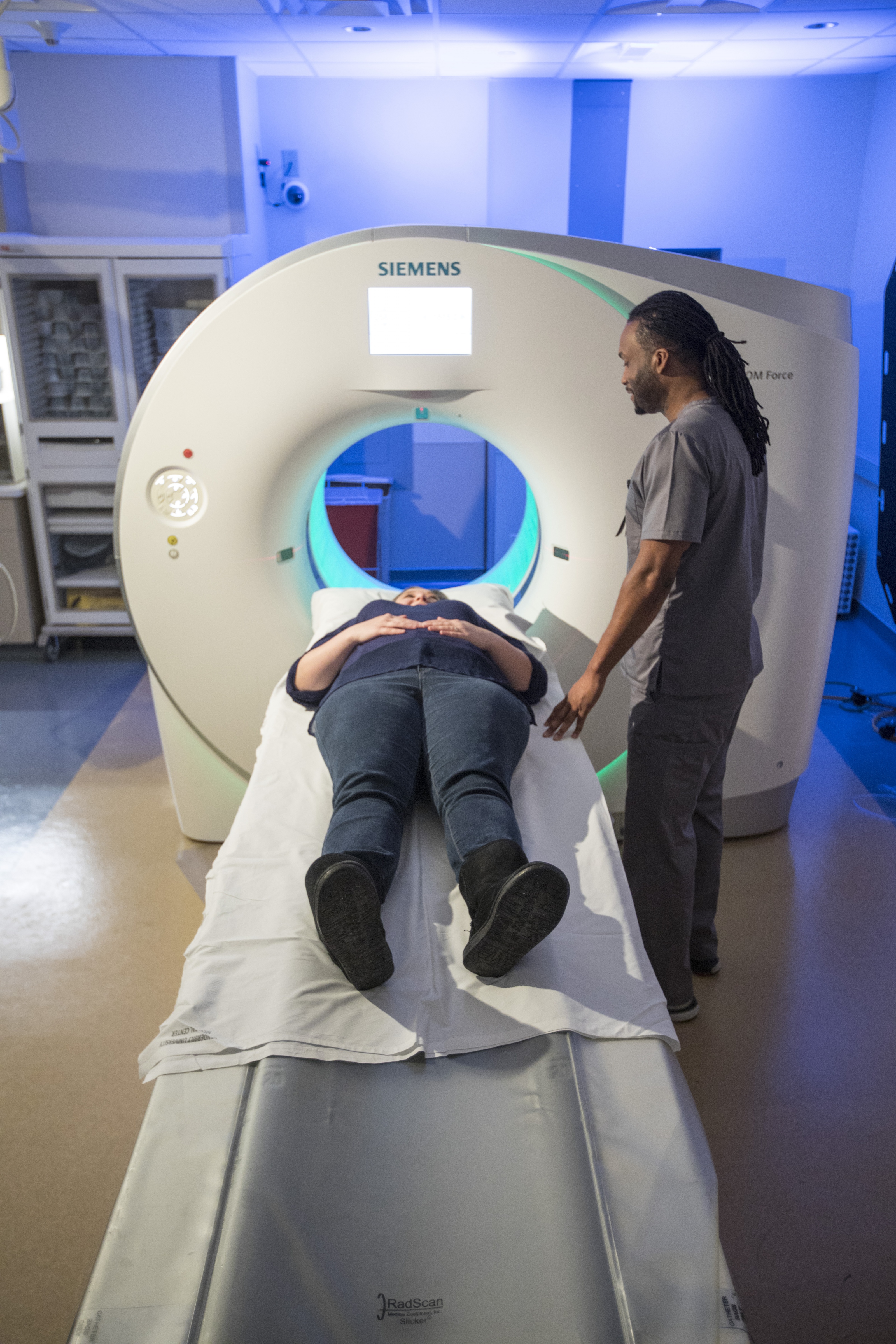
The first clinically operational in Tennessee, the Siemens Somatom Force CT scanners can create 384 slices per rotation, freeze motion in a beating heart, scan an entire patient from head-to-toe in under two seconds and image with multiple energies.
“These new super-fast CT units are a great benefit to patients and the professionals who operate them,” said Rich Pierce, Chief of Finance and Administration, Department of Radiology. “Worrying about a health concern that requires a diagnostic CT is never pleasant. Providing fast and precise imaging will help ease the burden.”
The dual-source, dual energy, wide detector CT systems reduce the radiation exposure and require lower amounts of intravenous contrast while producing higher quality exams. The Force CT scanner effectively contains two scanners in the same enclosure, which allows unparalleled options for completing an exam. One rotation of the gantry takes 0.25 seconds and each image has a temporal resolution of 66-msec.
Jeff Carr, M.D., Professor and Cornelius Vanderbilt Chair, Radiology and Radiological Sciences, says the ability to scan with two energies at the same time allows for precise tissue characterization and the ability to remove bones and metal artifacts that can obscure imaging of critical structures.
The scanner is also able to freeze motion, which allows imaging of small structures at a higher resolution. Moving structures that would previously be unrecognizable on an older CT scanner are also produced with better clarity.
“These new capabilities enhance the images we can produce, the types of studies we can perform and improve the care we provide to each and every patient,” said Dr. Carr. “The scanners will support the care of all patients by providing the most advanced imaging with the lowest possible exposure to X-rays and reduced intravenous contrast media.”
“Obtaining the new CT scanners was a team endeavor under the leadership of Jeff Balser, M.D., CEO, Mitch Edgeworth, CEO of Vanderbilt University Hospital, and Reed Omary, M.D., M.S., Professor and Chair, Department of Radiology and Radiological Sciences, who recognized the importance of this new, cutting-edge technology for our patients and the VUMC community,” added Dr. Carr.
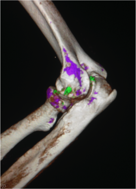
Elbow Arthritis Imaging – The new CT scanners provide not only high resolution imaging of the bones (3D) but also identify the chemistry, in this case the specific crystals seen in gouty arthritis are colored green.
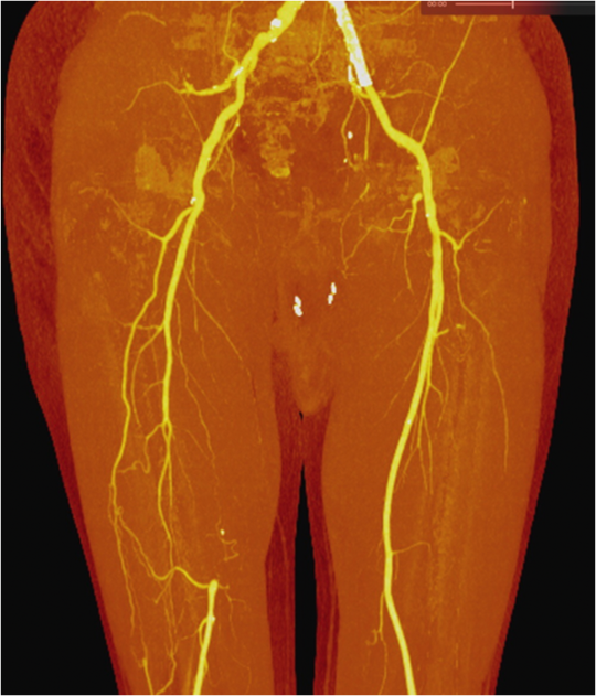
Examinations of the blood flow to the leg can be performed with both high resolution and lower amounts of intravenous contrast media, which is important for patients with reduced kidney function.