MD Imam Uddin, Ph.D. 
Assistant Professor of Ophthalmology and Visual Sciences
Vanderbilt University School of Medicine
Assistant Professor of Biomedical Engineering
Vanderbilt University School of Engineering
Office Address:
AA1324 Medical Center North
Vanderbilt University Medical Center
Nashville, TN 37232-8808
Office phone: 615-936-1447
email: md.i.uddin@vumc.org
Lab Address:
AA1309 & AA1315 Medical Center North
Vanderbilt University Medical Center
1161 21st Av S. Nashville, TN 37232-8808
phone: 615-936-9165 (AA1309), & 615-936-9181 (AA1315).
ORC ID: orcid.org/0000-0001-5611-9666
Lab website: https://lab.vanderbilt.edu/imam-uddin-lab/about/
Dr. Uddin is a primary faculty member (Tenure-track) in the Department of Ophthalmology and Visual Sciences, Vanderbilt University School of Medicine and has a secondary appointment in the Department of Biomedical Engineering. His long-term goal is to develop multi-modality imaging methods for retinal vascular diseases, primarily diabetic retinopathy (DR), neovascular or “wet” AMD, retinopathy of prematurity (ROP) and retinal vein occlusion (RVO). He has authored several publications in the field of molecular imaging with applications toward retinal vascular disease, holds several patents related to this area of research, and actively engaged in research toward clinical translation of these new technologies. Among many imaging methods, Dr. Uddin has developed HYPOX-4 for molecular imaging of retinal hypoxia in living tissues. As the PI on several Foundation- and NIH-funded projects, Dr. Uddin has laid the groundwork for trainees including early career T and K scholars and committed to diversity, equity and inclusion under his direct supervision.
Publications related to Project 1: Development of novel in vivo molecular imaging probes for retinal hypoxia– In a series of publications, we demonstrated the development of in vivo molecular imaging probes for retinal hypoxia. These publications have led to discover highly sensitive molecular imaging probes for retinal hypoxia, HYPOX-4 a water soluble, non-toxic hypoxia sensitive imaging agent. This imaging method will be useful in visualizing disease onset, progression and response to therapy in living retinal tissues.
a) Uddin, M. I.; Jayagopal, A.; McCollum, G. W.; Rong, Y.; Penn, J. S. In Vivo Imaging of Retinal Hypoxia using HYPOX-4-dependent Fluorescence in a Mouse Model of Laser-induced Retinal Vein Occlusion (RVO). Invest. Ophthalmol. Vis. Sci., 2017, 58(9), 3818-3824.
b) Uddin M.I., Jayagopal, A. (2020) Emerging Therapeutic Modalities for Diabetic Eye Disease. Topics in Medicinal Chemistry. Vol.35, page 161-187.
c) Uddin, M. I.; Evans, S. M.; Craft, J. R.; Capozzi, M. E.; McCollum, G. W.; Yang, R.; Marnett, L. J.; Uddin, M. J.; Jayagopal, A.; Penn, J. S. In Vivo Imaging of Retinal Hypoxia in a Model of Oxygen-Induced Retinopathy. Scientific Reports, 2016, 6, 31011.
d) Uddin, M. I., Evans, S. M., Craft, J. R., Marnett, L. J., Uddin, M. J. Joyagopal, A. Applications of azo-based probes for imaging retinal hypoxia. ACS Medicinal Chemistry Letters, 2015, 6 (4), 445-449.
e) Evans, S. M.; Kim, K.; Moore, C. E.; Uddin, M. I.; Capozzi, M. E.; Craft, J. R.; Sulikowski, G. A.; J. Joyagopal, A. Molecular Probes for Imaging of Hypoxia in the Retina. Bioconjugate Chem., 2014, 25 (11), 2030–2037.
PATENT (Number US10695446), Uddin, M. I.; Jayagopal, A.; Uddin, M.J.; Penn, J.S.; Marnett, L.J. (issue date: June 2020) Compositions and Method for Detecting Hypoxia. World Intellectual Property Organization, WO 2016/179117 A1.
Publications related to Project 2: Novel methods for in vivo molecular imaging of mRNA expression in the retina– Our efforts to develop novel in vivo molecular imaging probes have focused on visualizing mRNA in living retinal tissues. This effort yielded publications demonstrating the characteristics of functionalized gold nanoparticles targeting specific mRNA in living retinal cells and tissues.
a. Uddin, M. I.; Kilburn, T.C.; Duvall, L.C.; Penn, J.S. A novel method for visualizing and tracking endogenous mRNA in a specific cell population in pathological neovascularization. Nature Sci Rep, (In Revision) 2020.
b. Uddin, M. I.; Jayagopal, A.; Wong, A.; McCollum, G. W.; Wright, D. W.; Penn, J. S. Real-time imaging of VCAM-1 mRNA in TNF-α activated retinal microvascular endothelial cells using antisense hairpin-DNA functionalized gold nanoparticles. Nanomedicine:NBM, 2018, 14(1), 63-71.
c. Uddin, M. I.; Kilburn, T.C.; Rong, Y.; McCollum, G.W.; Wright, D.W.; Penn, J.S. Targeted imaging of VCAM-1 mRNA in a mouse model of laser-induced choroidal neovascularization (LCNV) using antisense hairpin-DNA functionalized gold-nanoparticles. Mol Pharm, 2018, 15(12), 5514-5520.
d. Uddin, M. I.; Kilburn, T.C.; Duvall, L.C.; Penn, J.S. Visualizing HIF-1alpha mRNA in a sub-population of bone marrow derived cells to predict retinal neovascularization. ACS Chemical Biology, (In Press) 2020.
PATENT (assigned), Uddin, M. I.; Penn, J.S.; Duvall, CL. PROBES FOR DETECTING RNA AND METHODS OF USE THEREOF. United States Patent Application, 2019.
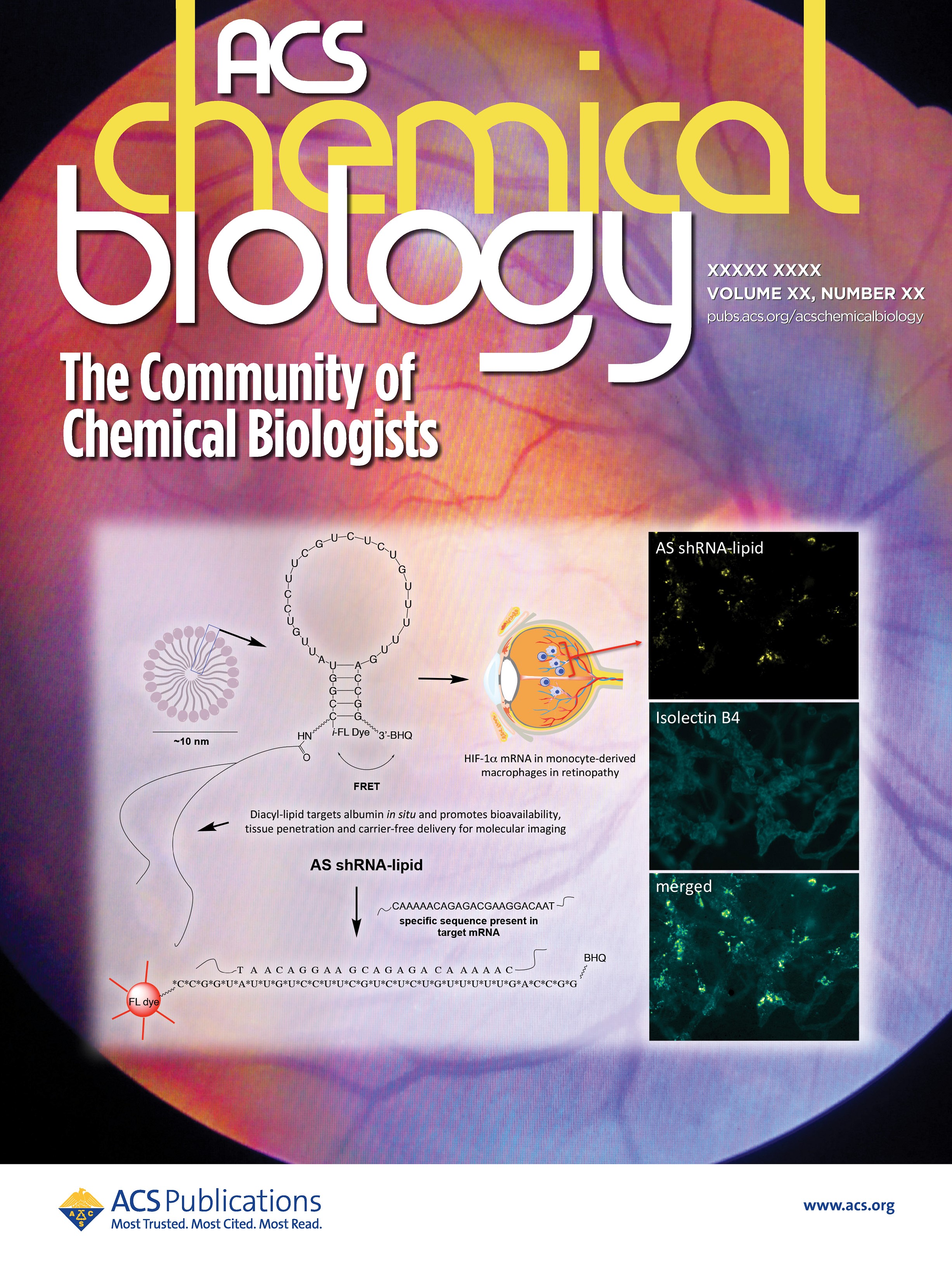
About the Image:
Selective visualization of monocyte-derived macrophages could be a powerful method to predict the onset and progression of ocular-angiogenesis at molecular level. In this image Dr. Uddin and colleagues described the synthesis of a new hybrid nanoparticle to visualize HIF-1 alpha mRNA selectively in monocyte-derived macrophages in a mouse model of ocular-angiogenesis.