For my Vanderbilt Vision Research Center page, please visit http://www.psy.vanderbilt.edu/faculty/calkinslab/
Overview
Our laboratory focuses on the mechanisms of neurodegeneration in glaucoma, the leading cause of irreversible blindness worldwide. Glaucoma blinds through the gradual loss of retinal ganglion cell neurons and their axons, which comprise the optic nerve. The loss of these neurons affects a major portion of the brain, which receives signals from the retina. Insight into the early molecular cascades involved in the degenerative process hopefully will lend insight into the identification of novel therapeutic targets. The laboratory uses systems, cellular and molecular approaches to investigate how risk factors in glaucoma, such as age and elevated ocular pressure, contribute to neurodegeneration in the disease and to test new treatments.
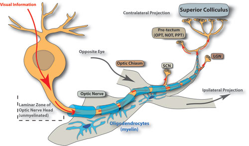
The axon of the retinal ganglion cell neuron projects to various nuclei of the brain through the optic nerve. In glaucoma, these these projections degenerate in response to key risk factors like aging and elevated ocular pressure.
Investigations
Calcium and Neurodegeneration. Recent work in the laboratory demonstrates that calcium plays a role early in the pathogenesis of the disease. We have found that retinal ganglion cells express numerous subunits of the transient receptor potential (TRP) family of cation channels that are characterized by a robust calcium conductance. In particular, the TRPV1 subunit in ganglion cells can be activated by pressure and contributes to increased intracellular calcium and ganglion cell death in isolation. By quenching this activation, stress due to pressure can be mitigated. Ongoing studies suggest that TRPV1 interacts with other TRP subunits and diverse transmembrane proteins to help regulate the flow of calcium in the ganglion cell body and axon.
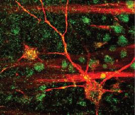
Retinal ganglion cell (red) expressing the TRPV1 channel (green) at specific locations (yellow). See papers by Sappington & Calkins (2008) and Sappington et al. (2009) listed below for details.
Axonal Transport and Neurodegeneration. Neurodegenerative disorders of the brain often entail early loss of transport along axonal tracts. Our work demonstrates that this is so also in glaucoma, where age and ocular pressure contribute to diminished transport of nutrients and other basic building blocks of neuronal function along the optic nerve. We have found that depleted transport is in fact among the earliest signs of pathology in glaucoma.
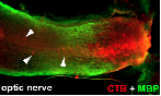
Retinal ganglion cell axons (green) in the optic nerve only partially transport dyes out of the retina (red) even at early stages in glaucoma. Arrows mark points of blocked transport in the nerve.
Select Recent Publications
- Sappington, R.M., Sidorova, T., Long, D.J. & Calkins, D.J. (2009) TRPV1: Contribution to retinal ganglion cell apoptosis and increased intracellular Ca2+ with exposure to hydrostatic pressure (Inv. Ophthal. Vis. Sci., 50, 717-728).
- Sappington, R.M. & Calkins, D.J. (2008) Pressure-induced translocation of NFkB and IL-6 release in retinal microglia involves Ca2+ and TRPV1 (Inv. Opthal. Vis. Sci., 49, 3004-3017).
- Wax, M.B., Tezel, G., Yang, J., Peng, G., Patil, R.V., Agarwal, N., Sappington, R.M., Calkins, D.J. (2008) Induced autoimmunity to heat shock proteins elicits glaucomatous loss of retinal ganglion cell neurons via activated T cell-derived Fas-ligand (J. Neurosci., 28, 12085-12096).
- Calkins, D.J., Horner, P.J., Roberts, R., Gradianu, M. & Berkowitz, B.A. (2008) Manganese-enhanced MRI of the DBA/2J mouse model of hereditary glaucoma (Inv. Ophthal. Vis. Sci., 49, 5083-5088.).
- Inman, D.M., Sappington, R.M., Horner, P.J. & Calkins, D.J. (2006) Quantitative correlation of optic nerve pathology with ocular pressure and corneal thickness in the DBA/2 mouse model of glaucoma (Inv. Opthal. Vis. Sci. 47, pp. 986-996).