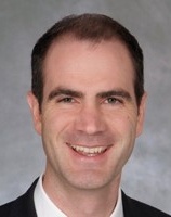
Changho Choi, Ph.D.
Optimization of spectrally-selective 180o RF pulse timings in J-difference editing (MEGA) of lactate.
Ganji SK, An Z, Tiwari V, Chang Y, Patel TR, Maher EA, Choi C.
Magnetic Resonance in Medicine 2022;87(3):1150-1164.
Glycine by MR spectroscopy is an imaging biomarker of glioma aggressiveness. Neuro-oncology.
Tiwari V, Daoud EV, Hatanpaa KJ, Gao A, Zhang S, An Z, Ganji SK, Raisanen JM, Lewis CM, Askari P, Baxter J, Levy M, Dimitrov I, Thomas BP, Pinho MC, Madden CJ, Pan E, Patel TR, DeBerardinis RJ, Sherry AD, Mickey BE, Malloy CR, Maher EA, Choi C.
Neuro-Oncology 2020;22(7):1018-1029.
3D high-resolution imaging of 2-hydroxyglutarate in glioma patients using DRAG-EPSI at 3T in vivo.
An Z, Tiwari V, Baxter J, Levy M, Hatanpaa KJ, Pan E, Maher EA, Patel TR, Mickey BE, Choi C.
Magnetic Resonance in Medicine 2019;81(2):795-802.
Distinction of the GABA 2.29 ppm Resonance using Triple Refocusing at 3T In Vivo
Tiwari V, An Z, Wang Y, Choi C.
Magnetic Resonance in Medicine 2018;80(4):1307-1319
Prospective longitudinal analysis of 2-hydroxyglutarate MR spectroscopy identifies broad clinical utility for the management of IDH-mutant glioma patients.
Choi C, Raisanen JM, Ganji SK, Zhang S, McNeil SS, An Z, Madan A, Hatanpaa KJ, Vemireddy V, Sheppard CA, Oliver D, Hulsey KM, Tiwari V, Mashimo T, Battiste J, Barnett S, Madden CJ, Patel TV, Pan E, Malloy CR, Mickey BE, Bachoo RM, Maher EM.
Journal of Clinical Oncology. 2016;34(33):4030-4039.
2-Hydroxyglutarate detection by magnetic resonance spectroscopy in IDH-mutated glioma patients.
Choi C, Ganji SK, DeBerardinis RJ, Hatanpaa KJ, Rakheja D, Kovacs Z, Yang XL, Mashimo T, Raisanen JM, Marin-Valencia I, Pascual JM, Madden CJ, Mickey BE, Malloy CM, Bachoo R, Maher EA.
Nature Medicine. 2012;18(4):624-629.
My research is focused on development of magnetic resonance spectroscopy (MRS) and identification of metabolic imaging biomarkers in brain cancers. Technical development is performed with tailoring of traditional MRS sequences, triple-refocusing MRS, and J-difference editing at 3T and 7T, along with high-resolution 3D spectroscopic imaging. Metabolites of interest include 2-hydroxyglurate (2HG), glycine, GABA, glutamate, glutamine, glutathione, lactate, etc.
We carry out or plan to conduct several projects in collaboration with clinicians. 1) Translation of MRS into clinics. A brain tumor MRS pipeline can enable clinicians to non-invasively assess the cellular and genetic alterations in brain cancers. 2) Brain cancer biomarker study. We examine association of metabolite levels with cancer cell proliferation, treatment response, and clinical outcomes. 3) Real-time metabolism study. We aim to identify the sources of metabolic alterations in brain tumors by measuring the changes in 1H MRS signals following administration of 13C labeled substrates in patients. 4) Deep-leaning MRS. We aim to build up a fast MRS analysis algorithm that can provide user-independent quantification of metabolites in gliomas. 5) MRS in neuro-psychiatric disorders. We use optimized MRS to identify metabolic alterations in schizophrenia, asthma, and depression.
Jeffry Nyman, Ph.D.
Nyman, J.S.*, Gorochow, L. E., Horch, R.A., Uppuganti, S., Zein-Sabatto, A., Manhard, M.K. and M.D. Does. Partial removal of pore and loosely bound water by low-energy drying decreases cortical bone toughness in young and old donors. Journal of the Mechanical Behavior of Biomedical Materials. In press. (http://www.sciencedirect.com/science/article/pii/S1751616112002263)
Lian, N., Lin, T., Liu, W., Wang, W., Li, L., Sun, S., Nyman, J.S. and X. Yang. Transforming Growth Factor �?² suppresses osteoblast differentiation via vimentin activating transcription factor 4 (ATF4) axis. The Journal of Biological Chemistry. 287:35975-84, 2012.
O'Neill, K.R., Stutz, C.M., Mignemi, N.A., Burns, M.C., Murry, M.R. Nyman, J.S., and J.G. Schoenecker. Micro-computed tomography assessment of the progression of fracture healing in mice bone. Bone. 50:1357-67, 2012.
Horch, R.A., Gochberg, D.F., Nyman, J.S., and M.D. Does. Clinically-compatible MRI strategies for discriminating bound and pore water in cortical bone. Magnetic Resonance in Medicine. 68:1774-84, 2012.
Nyman, J.S.*, Makowski, A.J., Patil, C.A., Masui, T.P., O'Quinn, E.C., Bi, X., Guelcher, S.A., Nicollela, D.P. and A. Mahadevan-Jansen. Measuring differences in compositional properties of bone tissue by confocal Raman Spectroscopy. Calcified Tissue International. 89:111-22, 2011.
The goal of my research is to lower the number of bone fractures associated with osteoporosis, diabetes, cancer, and aging.
One project investigates the determinants of bound water in bone and the ability of 1H Nuclear Magnetic Resonance to explain the age- and diabetes-related decrease in bone's resistance to fracture. Another project aims to identify novel Raman spectrocospy signatures that can distinguish normal bone quality from poor bone quality. Using wild-type and genetically modified mice with and without exogenous treatments, I also have several projects investigating how advanced glycation end-products, matrix proteins, and growth factors affect bone toughness. In collaboration with my colleagues at the Center for Bone Biology, we test new therapeutics to prevent fractures or heal fractures in pre-clinical rodent models of disease (namely, diabetes, bone metastasis, neurofibromatosis type 1).
Yu Zhao
Postdoctoral Fellow 2019 thru 022
Current: Associate professor at West China Hospital
Susan Meyn
In my role in the Office of Research (OOR), I support the research and education missions of the Medical Center overall. This work is extended through focus on centers and institutes such as VUIIS. For those interested in the broader mission of the OOR, please visit our website here.
My current emphasis involves supporting the Director of VUIIS in overseeing the administration and financial health of the Institute, through collaborative efforts to maximize existing resources and identify new opportunities to sustain and grow the VUIIS research and training missions.
Simon Vandekar
Alexander-Bloch, A.F., Shou, H., Liu, S., Satterthwaite, T.D., Glahn, D.C., Shinohara, R.T., Vandekar, S.N., Raznahan, A., 2018. On testing for spatial correspondence between maps of human brain structure and function. NeuroImage 178, 540?551. doi: 0.1016/j.neuroimage.2018.05.070
Vandekar, S.N., Reiss, P.T., Shinohara, R.T., 2018a. Interpretable High-Dimensional Inference Via Score Projection With an Application in Neuroimaging. Journal of the American Statistical Association 0, 1?11. doi: 10.1080/01621459.2018.1448826
Vandekar, S.N., Satterthwaite, T.D., Rosen, A., Ciric, R., Roalf, D.R., Ruparel, K., Gur, R.C., Gur, R.E., Shinohara, R.T., 2017. Faster family-wise error control for neuroimaging with a parametric bootstrap. Biostatistics. doi: 10.1093/biostatistics/kxx051
Vandekar, S.N., Satterthwaite, T.D., Xia, C.H., Ruparel, K., Gur, R.C., Gur, R.E., Shinohara, R.T., 2018b. Robust Spatial Extent Inference with a Semiparametric Bootstrap Joint Testing Procedure. arXiv:1808.07449 [stat].
Vandekar, S.N., Shinohara, R.T., Raznahan, A., Roalf, D.R., Ross, M., DeLeo, N., Ruparel, K., Verma, R., Wolf, D.H., Gur, R.C., Gur, R.E., Satterthwaite, T.D., 2015. Topologically Dissociable Patterns of Development of the Human Cerebral Cortex. J. Neurosci. 35, 599?609. 10.1523/JNEUROSCI.3628-14.2015
My interests pertain to statistical issues in medical image analysis such as quantification of uncertainty and replicability and performing high dimensional inference.
My work lately has focused on making accurate probabilistic statements for high-dimensional neuroimaging data such as computing voxel-wise and spatial extent adjusted p-values that describe how unlikely the observed results are under a particular null hypothesis. In imaging this requires dealing with low-rank estimates for complex covariance matrices, issues of normal approximations in high-dimensional data, and model misspecification. I developed several new (semi)parametric bootstrap joint (PBJ) inference procedures, which are available as an R package (pbj) on github. My current research is attempts to accurately estimate the probability that a thresholded statistical image contains all truly activated regions using small sample sizes. This is more akin to constructing confidence intervals than hypothesis testing and affords more power when the null hypothesis is false. I am also interested in the estimation of the proportion of the image with a nonzero effect size, inference for machine learning models of imaging data, topological data analysis, and unsupervised deep learning.
Kenny Tao
Malone, J. D., El-Haddad, M. T., Bozic, I., Tye, L. A., Majeau, L., Godbout, N., Rollins, A. M., Boudoux, C., Joos, K. M., Patel, S. N., and Tao, Y. K., "Simultaneous multimodal ophthalmic imaging using swept-source spectrally encoded scanning laser ophthalmoscopy and optical coherence tomography," Biomed. Opt. Express 8, 193-206 (2017)
Tao, Y. K., Srivastava, S. K., and Ehlers, J. P., "Microscope-integrated intraoperative oct with electrically tunable focus and heads-up display for imaging of ophthalmic surgical maneuvers," Biomedical optics express (Featured Cover) 5, 1877-1885 (2014).
Ehlers, J. P., Srivastava, S. K., Feiler, D., Noonan, A. I., Rollins, A. M., and Tao, Y. K., "Integrative advances for oct-guided ophthalmic surgery and intraoperative oct: Microscope integration, surgical instrumentation, and heads-up display surgeon feedback," PloS one 9, e105224 (2014).
Tao, Y. K., Shen, D., Sheikine, Y., Ahsen, O. O., Wang, H. H., Schmolze, D. B., Johnson, N. B., Brooker, J. S., Cable, A. E., Connolly, J. L., and Fujimoto, J. G., "Assessment of breast pathologies using nonlinear microscopy," Proceedings of the National Academy of Sciences of the United States of America 111, 15304-15309 (2014).
Tao, Y. K., Kennedy, K. M., and Izatt, J. A., "Velocity-resolved 3d retinal microvessel imaging using single-pass flow imaging spectral domain optical coherence tomography," Optics express 17, 4177-4188 (2009).
My lab develops novel optical imaging systems for clinical diagnostics and therapeutic monitoring in ophthalmology, gastroenterology, and oncology.
Biomedical optics enable noninvasive subcellular visualization of tissue morphology, biological dynamics, and disease pathogenesis. Ongoing research in my lab focuses on clinical translation of therapeutic tools for image-guided intraoperative feedback using modalities including optical coherence tomography and nonlinear microscopy. My lab has also developed optical imaging techniques that exploit intrinsic functional contrast for in vivo monitoring of blood flow and oxygenation as surrogate biomarkers of cellular metabolism and early indicators of disease. The majority of my research involves multidisciplinary collaborations between investigators in engineering, basic sciences, and medicine.
Ryan Robison, Ph.D
As a Philips Clinical Scientist, I collaborate with VUMC faculty and researchers. Current projects include robust axial T2* and diffusion imaging of the spinal cord, spiral methods for sodium imaging, MR spectroscopy of lactate, and oscillating gradient spin echo diffusion.
My research interests include advanced MRI acquisition methods, data reconstruction, and artifact corrections. While all MR imaging research is of interest to me, I have a particular interest in brain and spinal cord imaging.


