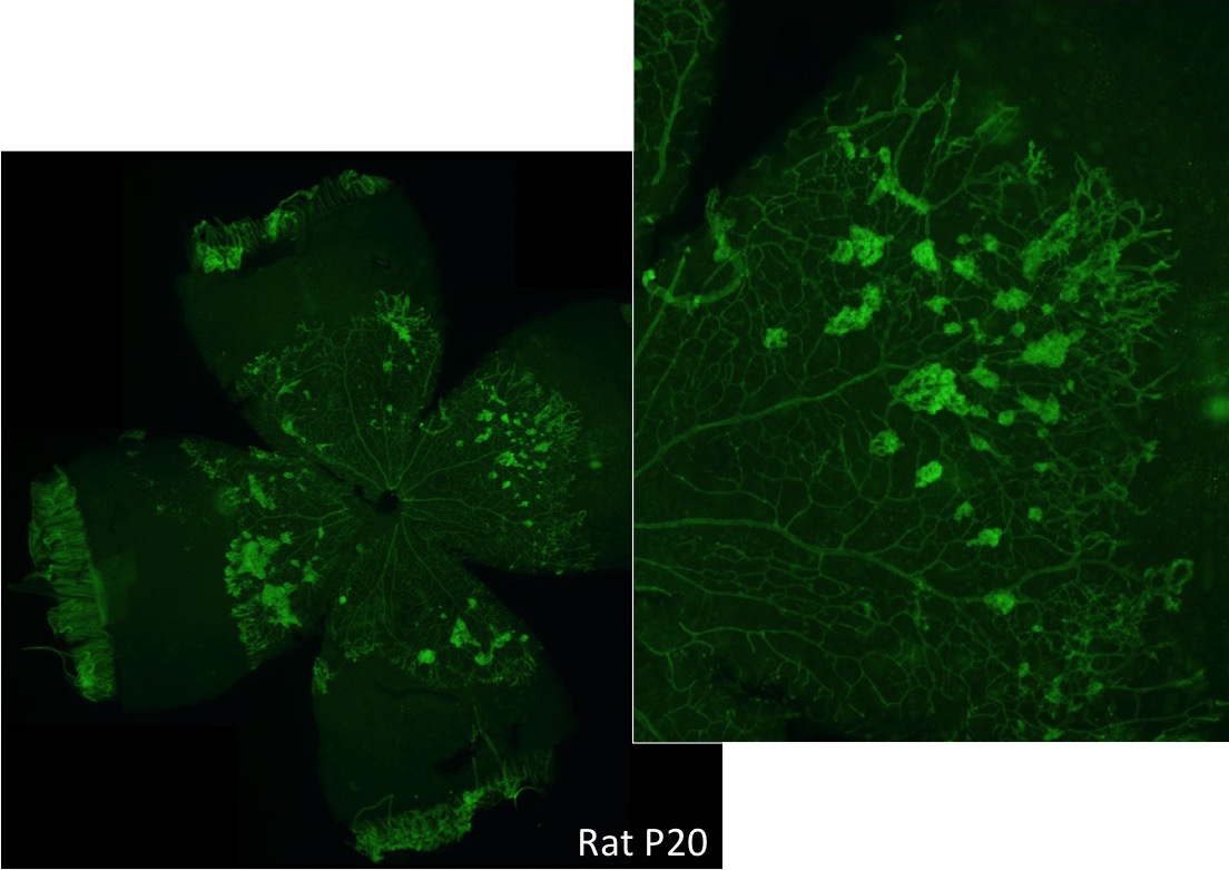
Neovascularization Assessment
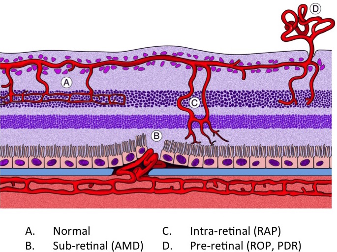
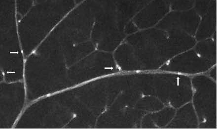
Neovascular area can be assessed for both the OIR and LCNV models. We carefully dissect the retina or choroid, stain for blood vessels, and flat-mount the tissue. We then measure neovascular growth using computer-assisted image analysis tools. Our methods allow for precise and highly reproduceable data.
Vascular Permeability
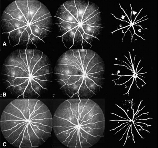
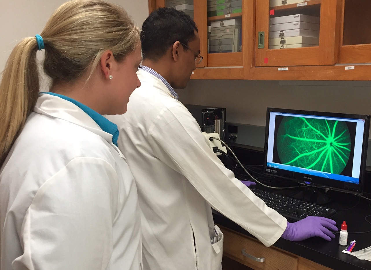
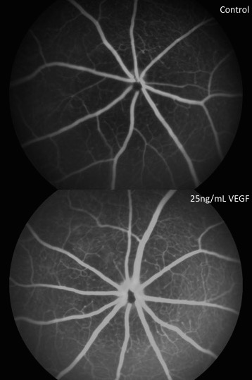
Increased vascular permeability is a damaging component of diabetic retinopathy. To test whether a drug has an effect on permeability, we use one or more of several methods. We measure the extravasation of Evan's blue dye or fluorescein-conjugated dextrans from the vasculature into the retinal tissue, we examine fluorescein angiography, or we measure retinal thickness via OCT.
Leukostasis
Leukostasis, the adherence of leukocytes to endothelium, is a major component of non-proliferative diabetic retinopathy and is a general indicator of tissue inflammation. We measure retinal leukostasis by infusing animals with fluorescein-conjugated concanavalin A, flushing the circulation with buffer perfusion, carefully dissecting and flat-mounting retinas, and counting the number of leukocytes remaining in the lumenal space of blood vessels.
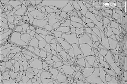
Trypsin Digest
Diabetic retinopathy is characterized by an unhealthy vasculature with loss of pericyte and endothelial cells. Trypsin digests can be used to carefully extract only the retinal vasculature to assess its characteristics. Once extracted and flat-mounted, the pericyte to endothelial cell ratio can be counted, the number of ghost capillaries can be determined, or immunohistochemistry can be performed.