Sun
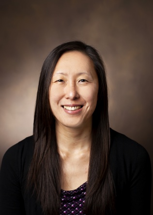
H.
Peck
Ph.D.
Medicine, Division of Clinical Pharmacology
Biochemistry, Vanderbilt University School of Medicine
Biomedical Engineering, Vanderbilt University School of Engineering
Research Health Scientist
Nashville Veterans Affairs Medical Center
Background
Dr. Peck received her doctorate in Chemical Biology from Harvard University under the mentorship of Dr. David Liu in the Department of Chemistry and Chemical Biology, where she developed novel molecular evolution techniques to study protein-protein interactions and protein function. Dr. Peck’s interests in macromolecular chemistry and related biological functions led her to pursue postdoctoral training in lysosomal storage disorders and pathological consequences of dysregulated metabolism, first in the Division of Biology at Kansas State University under the mentorship of Dr. Stella Lee, elucidating the role of N-glycosylation in protein trafficking, folding, and function in neuronal ceroid lipofuscinoses, a family of neurodegenerative lysosomal storage disorders. She continued her postdoctoral training in the Department of Orthopaedic Surgery at the University of Pennsylvania as an NIH F32 postdoctoral fellow under the mentorship of Drs. Lachlan Smith and Eileen Shore. Under this award, Dr. Peck investigated the molecular mechanisms underlying dysregulated vertebral bone development in mucopolysaccharidoses, a family of lysosomal storage disorders that presents with severe musculoskeletal deformities as a consequence of aberrant glycosaminoglycan metabolism. During her postdoctoral fellowship, Dr. Peck also elucidated developmental signals underlying the formation of the glycosaminoglycan-rich region of the intervertebral disc known as the nucleus pulposus and led studies into cell-based regenerative therapies for treating disc degeneration.
Dr. Peck’s overall research interests are to combine her chemistry background with her postdoctoral experience in musculoskeletal biology to investigate carbohydrate-mediated signaling in spine development and homeostasis, how dysfunction in these signals lead to degeneration and diseased states, and to develop biologic and engineering approaches to regenerative therapies for musculoskeletal tissues.
Lab Address
Medical Research Building IV / Light Hall
2215B Garland Ave
Nashville
Tennessee
37232
The overarching focus of my research laboratory is understanding the role of the extracellular matrix (ECM) in musculoskeletal biology, particularly of carbohydrate-mediated signaling and differentiation as regulated by glycosaminoglycans (GAGs). Glycosaminoglycans are unbranched polysaccharides that are abundantly present in many organ systems in the body. In musculoskeletal tissues, GAGs are typically thought to serve primarily structural roles, as they are highly polar, hydrophilic moieties capable of high osmotic swelling pressure. However, there is mounting evidence that GAGs in the ECM interact with secreted signaling molecules, cells, and other ECM components, and these interactions play a substantial role in controlling the activity of resident cells and subsequent tissue development, function, and homeostasis. While the primary molecules of interest are GAGs, our research encompasses the investigation of related ECM components such as collagens and growth factors that interact with GAGs to carry out important biological functions. We combine analytical chemistry, biochemistry, and molecular biology methods to identify the chemical composition of the ECM and how particular components regulate development and homeostasis, as well as how dysregulated ECM deposition or degradation contribute to disease or degeneration. Ultimately, we seek to understand these mechanisms in order to develop regenerative therapies for bone, intervertebral disc, and cartilage. To this end, we have multiple ongoing projects in the lab that are designed to elucidate normal and pathologic contributions of the ECM in musculoskeletal tissues.
- Dysregulated extracellular matrix deposition in heterotopic ossification.
- Heterotopic ossification (HO) is the pathologic formation of bone in extra-skeletal tissues. HO can develop following significant trauma such as blast, brain, or spinal cord injuries, burns, or after routine surgical procedures such as joint replacement or amputation surgery. Ectopic bone formation leads to impaired wound healing, chronic infection, and chronic pain, which can hinder mobility and function as well as the use of prosthetics. This can lead to further related health complications such as opioid addiction, depression, and suicide. HO affects the Veteran population disproportionately but has been found in all patient populations. The molecular mechanisms that drive this aberrant bone formation are not well-understood, and thus, effective therapeutics are lacking. We are focused on elucidating the mechanisms underlying aberrant bone formation as driven by tissue and molecular level changes to the ECM.
- Extracellular matrix-mediated signaling and patterning in early spine development.
- The embryonic notochord is a unique, transient structure that serves as the main signaling center for directing developmental patterning. The notochord starts out as a GAG-rich rod-like structure, which over time becomes sequestered in the ECM-rich central region of the intervertebral disc referred to as the nucleus pulposus (NP). My previous work identified large changes in global expression profiles of ECM molecules during this notochord to NP transition. However, the molecular details of how this GAG-rich structure directs spine patterning and transforms into the NP in early embryonic development remains unknown. We are interested in identifying ECM-mediated signaling that drive spine formation.
- Extracellular matrix regeneration as a therapeutic strategy for back pain.
- The nucleus pulposus (NP) is the ECM-rich central region of the intervertebral disc that is responsible for distributing compressive loads on the spine. Pathological changes to this region are often thought to initiate progressive structural deterioration of the entire intervertebral joint. Degeneration of the disc is strongly implicated as a cause of low back pain, which ~85% of people will experience in their lifetime. This is a significant clinical problem that results in over $100 billion in healthcare and socioeconomic costs every year. Current treatments are mostly palliative, and the fraction of patients who are candidates for surgical interventions have high failure rates. Importantly, none of the available treatments maintain or restore native disc structure or biomechanical function, and thus, there is a significant clinical need to develop therapeutic approaches that manage symptoms as well as regenerate native disc tissue. We are working to elucidate the pathologic breakdown of the ECM that leads to intervertebral disc degeneration on the molecular level. Furthermore, in conjunction with our interests in identifying the ECM-mediated signaling that drives early NP formation, we are working on applying these developmental signals to induce ECM formation as a potential treatment option for disc degeneration.
- Pathological changes to extracellular matrix composition as a result of sepsis.
- Systemic inflammatory response syndrome, or sepsis, is a serious medical condition with a high mortality rate. Even following recovery, sepsis patients often suffer from a myriad of long-lasting medical complications, which include neurological dysfunction, cardiovascular disease, and musculoskeletal disability. Interestingly, the organ systems that are affected in post-sepsis syndrome are often not the site of the primary infection that leads to sepsis. In collaboration with Dr. Fiona Harrison (Division of Diabetes and Endocrinology and The Vanderbilt Brain Institute), we are seeking to understand the molecular connections between systemic inflammation and resulting secondary pathology by elucidating the changes that occur in ECM metabolism as a result of sepsis.
Other areas of interest in development:
- The role of extracellular matrix synthesis in bone formation and fracture healing.
- The effects of metabolic disorders (obesity, diabetes, etc.) on extracellular matrix synthesis in wound healing.
sun.peck@vumc.org
Postdoctoral Fellow, Department of Orthopaedic Surgery, University of Pennsylvania
Postdoctoral Fellow, Division of Biology, Kansas State University
Ph.D., Chemical Biology, Harvard University
M.S., Chemistry, University of Pennsylvania
B.A., Biochemistry and Music, University of Pennsylvania
