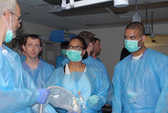
Under the instruction of body imaging specialists and faculty members Asma Ahmad, M.D., and Geoffrey Wile, M.D., first- and second-year radiology residents recently participated in a valuable hands-on workshop at the Vanderbilt University Medical Center’s (VUMC) cadaver laboratory.
The generously donated cadavers of a 96-year-old male and 94-year-old female provided residents with the opportunity to learn how to properly perform ultrasound (US)-guided liver and kidney biopsies, and how to treat tumors such as renal cell carcinoma with image-guided cryoablation.
“This hands-on workshop was an integrative experience for young physicians, teaching them how to perform complex US-guided interventions,” said Ahmad. “Our residents were able to become more familiar with the equipment, improve their hand-eye coordination and ask questions prior to interactions with patients.”
First-year radiology resident, Patrick Couture, M.D., says hands-on workshops such as these are an integral part of the medical education process because they allow students to work closely with faculty in order to practice procedures without the time or instructional constraints of actual procedures.
“Having multiple faculty members available to offer their tips and tricks proved invaluable,” added first-year radiology resident Brittany Burns, M.D. “The knowledge and understanding I obtained built confidence in providing procedure-based patient care, and I hope to do more of these workshops in the future!"