Islet and Pancreas Analysis (IPA) Core - Imaging
The IPA Core is thrilled to announce the launch of our new multiplexed imaging service! We hope this will be a valuable assay for core customers already using immunohistochemistry in their research and will also attract new customers within the Diabetes Center who study organs besides the pancreas (we can image these too!).
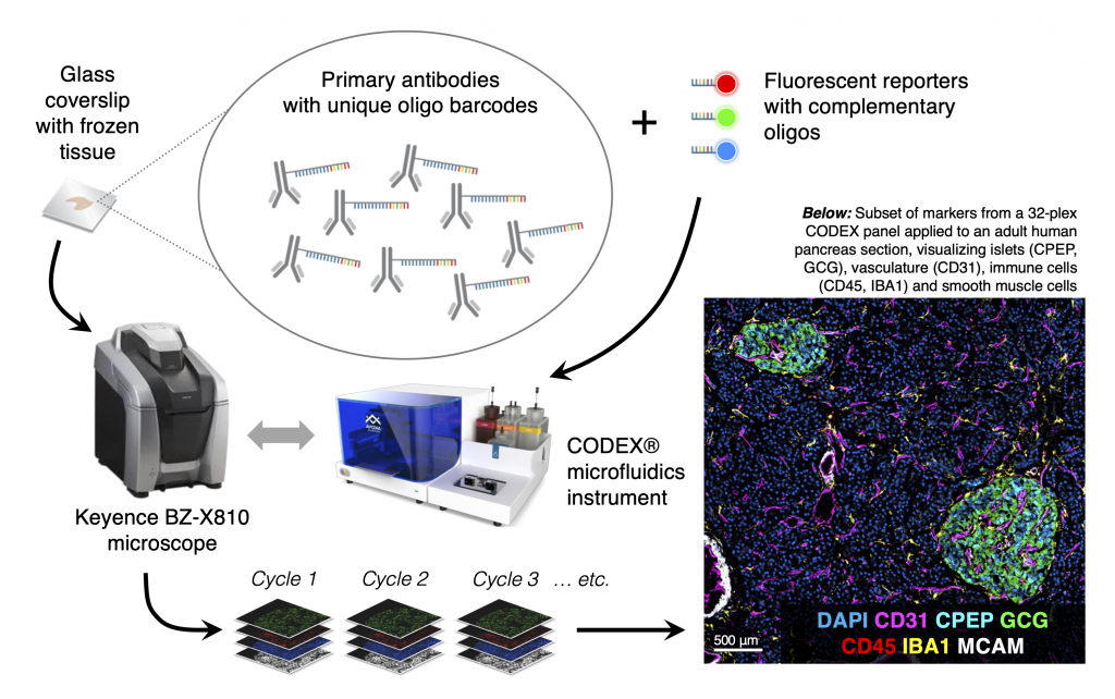
CO-Detection by indEXing (CODEX®; now branded as PhenoCycler™ by Akoya Biosciences) is an antibody- and fluorescence-based method of visualizing up to 40 antigens on a single tissue section. With each antibody conjugated to a unique oligonucleotide barcode, antigens are visualized by adding groups of fluorescently-conjugated complementary oligos and then merged to create a multi-layered image.
|
|
IPA will offer antibodies for all islet cell hormones, as well as Ki67 and vascular and ductal markers. Right now we are focusing on cryopreserved tissue samples (either flash frozen or lightly fixed in PFA) from human and mouse. Many immune antibodies are available off-the-shelf, so spleen and other tissues are also easily accommodated. Click here to explore CODEX images of human pancreas via Pancreatlas. To get started or learn more about initiating a project, e-mail Diane to set up a brief meeting.