Our research focuses on the use of non-conventional quantitative magnetic resonance imaging (MRI) techniques to untangle disease processes in Multiple Sclerosis (MS).
We use MRI at high and ultra-high field both in persons but also animals in which the model of the disease has been reproduced.
In 2019 the National MS Society (NMSS) has funded our lab to study the role of chronic inflammation in persons with MS, at the time of disease diagnosis. Specifically, our team is using ultra high field (7.0 Tesla) MRI to detect paramagnetic rim lesions in newly diagnosed persons and understand if their presence so early in the disease course is associated with a less favorable clinical outcome. Paramagnetic rim lesions are chronic plaques associated with a slow expansion over time and are considered a surrogate of chronic inflammation in MS.
In 2021 the VA Healthcare System has funded our lab to study neurodegeneration in early MS. We are using state-of-the art quantitative MRI methods to derive indirect estimates of myelin and axonal integrity and quantify the impact of neuroxonal injury early in the disease course. These methods include the selective inversion recovery quantitative magnetization transfer (SIR-qMT) and multi-compartment diffusion MRI with the spherical mean technique (SMT).
In 2021 the National Institutes of Health (NIH) has funded our lab to further study the pathological sensitivity and specificity of our SIR-qMT and SMT to myelin and axonal injury using two mice models of MS.
In 2024 NMSS funded our lab to study the accuracy of paramagnetic rim lesions detection using high and ultra-high filed susceptibility sensitive MRI.
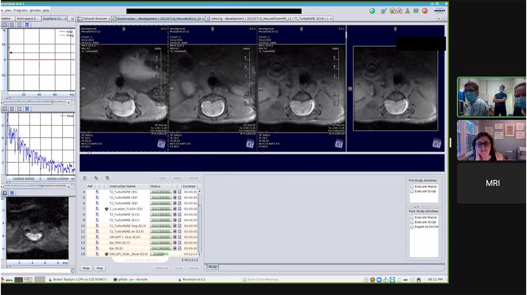
A nine-hour-long MRI session between Vanderbilt and Dartmouth University, Saturday July 16, 2022.