Staph and Osteoclasts

Staph
and
Osteoclasts

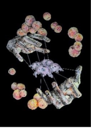
Staphylococcus aureus mediates immunological cross-talk between osteoclasts, cells that are responsible for degrading bone and remodeling the skeletal system. Staph found in infections can “manipulate” osteoclasts and promote bone damage and loss. 

2021. Digital media, reprinted. 
By Eddie Qian for the James Cassat laboratory

Multiplexed Molecular Imaging of Human Kidney

Multiplexed Molecular
Imaging of
Human Kidney

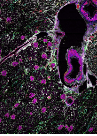
CODEX multiplexed immunofluorescence image of a human kidney tissue section. These data were collected as part of a multimodal imaging study integrating MALDI imaging mass spectrometry and CODEX to discover the relationships between lipid and metabolite distributions and cellular organization in donors with hypertension and type-2 diabetes for the Human Biomolecular Atlas Program (HuBMAP). 

By Allison Esselman, Thai Pham, and Melissa Farrow 
Spraggins Research Laboratory 
Department of Cell & Developmental Biology 
Mass Spectrometry Research Center 
Vanderbilt University School of Medicine

COX-2, Human Enzyme

COX-2,
Human Enzyme

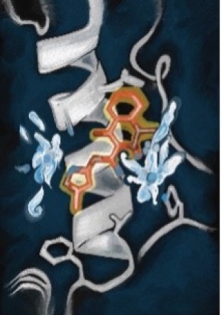
Larry Marnett, former Dean of Basic Sciences, was dedicated to his work with COX-2, a human enzyme that is targeted by non-steroidal anti-inflammatory drugs. This image is based on the structure of the NSAID isoxicam when bound to COX-2, a structure that Marnett’s lab determined in 2014. Water molecules mediate a crucial aspect of this interaction at amino acid residues R120 and Y385. Oxicams (including isoxicam) are the only class of NSAIDs for which water-mediated binding to COX-2 is observed. The structure, including the water molecules, is essential for explaining the structure-activity of this series of drugs. 

(Vestigo Cover, Issue 4) Illustration by Kendra H. Oliver.

Bacterial Flagellar Motor Assembly

Bacterial Flagellar
Motor Assembly

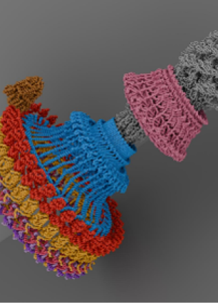
Bacterial Flagellar Motor Assembly: The Salmonella flagellar motor, which enables bacterial movement, is shown here with its key components: the C-ring proteins (FliG in red, FliM in yellow, and three FliN in shades of purple), the MS- ring (FliF in blue), the LP-ring (pink), the rod (gray), and the MotAB stator complex (brown). 

By Prashant Singh, Iverson Lab 
Vanderbilt University

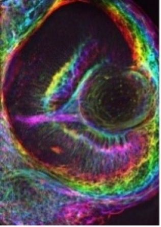
Zebrafish Eye

Zebrafish
Eye

The eye of a Zebrafish embryo at 72 hours post fertilization was imaged by spinning disk confocal microscopy. The embryo was labeled with Phalloidin-568 to label actin filaments to visualize the action cytoskeleton in the embryonic eye. The image is a projection of a 3D image with the rainbow colors showing the change of the signal through the Z dimension. 

Microscopist: Zachary Sanchez, PhD Candidate, Dylan Burnette Lab, Cell and Developmental Biology

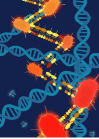
Acinetobacter baumannii DNA rearrangement

Acinetobacter
baumannii DNA
rearrangement

Mutations in the genome of A. baumannii increases A. baumannii's cell size and antimicrobial resistance, making it more harmful. This is shown in that the bacteria are a deeper red as they become more virulent. 

Artist Name: Alexa Marcus, work from the Skaar lab.

Microbe hunting in caves

Microbe
hunting in
caves

Brian Bachmann rappelling into a Tennessee cave to sample hypogean microbes for antibiotic and anticancer natural product discovery.

Photo by Jacob Burghart

CcmA Distribution Patterns on Heliobacter pylori

CcmA Distribution
Patterns on
Heliobacter pylori

This dynamic sculpture illustrates the 3D distribution of CcmA proteins on the cell wall surfaces of four H. pylori mutants (with each mutant type shown in a different color under natural light). When black light is introduced, the CcmA protein distribution on each cell becomes visible and begins to glow. Each cell shape is taken from a real cell in the lab, along with its corresponding protein distribution. 

2022. Digital Media, reprinted. 
By Mariana Smith, Ben Bratton Lab

Drosophila neural circuits

Drosophila
neural
circuits

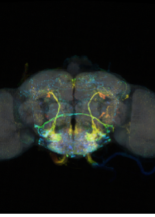
Sensory-motor neurons in central brain of Drosophila melanogaster

By: Emily Kophs, Ava Thorsen, Olivia Nunn, Suver Lab