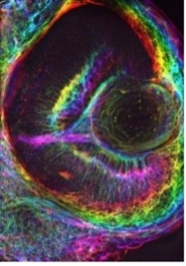
Zebrafish Eye
The eye of a Zebrafish embryo at 72 hours post fertilization was imaged by spinning disk confocal microscopy. The embryo was labeled with Phalloidin-568 to label actin filaments to visualize the action cytoskeleton in the embryonic eye. The image is a projection of a 3D image with the rainbow colors showing the change of the signal through the Z dimension.
Microscopist: Zachary Sanchez, PhD Candidate, Dylan Burnette Lab, Cell and Developmental Biology